Use the zoom controls to enlarge the algorithm, or open the full-screen view.

The algorithm could not load. Please reload the page.
Download the 5 × 8-inch PDF · Open the static algorithm
- Assessment, resuscitation, and cause-specific treatment proceed together
The Algorithm
01 ABCs / Initial Assessment
Assess Severity
-
Airway: Speech, stridor, swelling, secretions, and airway protection
-
Breathing: Rate / depth, tripoding, accessory muscles, retractions, paradoxical breathing, air entry, and asymmetry
-
Circulation / Consciousness: Pulse, BP, perfusion, and mental status
-
Impending Failure: Exhaustion, weakening effort, altered consciousness, or persistent hypoxemia
-
SpO₂ / Blood Gas: Monitor oxygenation; blood gas for suspected ventilatory failure—not saturation alone
Empiric Treatment / Respiratory Support
-
IV access / monitor / pulse oximetry; suction and airway equipment ready
-
Oxygen: Correct hypoxemia; target 88–92% when at risk of hypercapnic respiratory failure
-
Inadequate Breathing With A Definite Pulse: Open the airway and support ventilation with a bag-mask
-
Unresponsive / Absent Or Abnormal Breathing: No definite pulse within 10 seconds → CPR / defibrillator pathway
-
Noninvasive Support: Select by physiology; assess airway protection, aspiration risk, and hemodynamics
-
BiPAP: COPD with acute hypercapnic respiratory acidosis
-
CPAP / BiPAP: Cardiogenic pulmonary edema with respiratory failure
-
High-Flow Nasal Cannula (HFNC): Escalating hypoxemic respiratory failure, such as pneumonia
-
-
Shock: Cause-directed fluids / blood / vasopressors; reassess
Intubation / RSI
-
Failed oxygenation / ventilation, loss of airway protection, or anticipated deterioration requiring airway control
-
Preoxygenation / perfusion support / rescue airway ready; do not delay for repeated ineffective support
02 R/O Critical Causes
Airway Obstruction
-
Red Flags: Choking, stridor, inability to speak, weak cough, or absent airflow despite respiratory effort
-
Clear The Airway: Open / suction; remove a visible accessible object under direct visualization
-
Adult Choking: Effective cough → encourage coughing; severe obstruction while responsive → 5 back blows + 5 abdominal thrusts, repeated
-
Late pregnancy / unable to encircle abdomen → chest thrusts instead
-
Unresponsive → CPR starting with compressions; remove visible objects, no blind sweeps
-
-
Persistent obstruction → expert airway / bronchoscopy; cannot oxygenate → emergency airway rescue
Anaphylaxis
-
Red Flags: Abrupt respiratory compromise or hypotension after a likely trigger; hives / swelling may be absent
-
IM epinephrine immediately; repeat for persistent compromise
-
Airway support / oxygen / IV fluids for shock; refractory compromise → monitored epinephrine infusion
Angioedema
-
Red Flags: Progressive tongue / floor-of-mouth swelling, voice change, drooling, or stridor
-
Early expert airway management; surgical rescue ready
-
Possible anaphylaxis → IM epinephrine; ACE-inhibitor swelling → stop the ACE inhibitor; do not wait for allergy medications to restore the airway
Tension Pneumothorax
-
Red Flags: Sudden dyspnea, unilateral absent breath sounds, severe respiratory compromise / shock
-
Suspected tension + instability → immediate decompression, followed by chest drainage
-
Do not wait for imaging or tracheal deviation; bedside ultrasound only when it does not delay treatment
03 Evaluate Emergent Diagnoses
COPD / Asthma
-
Red Flags: Known obstructive disease, smoking / exposure history, wheezing, prolonged expiration, and reduced air entry
- Prior intubation / ICU admission, escalating reliever use, exhaustion, or a silent chest increase concern
-
Evaluation: Severity / SpO₂; blood gas for suspected ventilatory failure; CXR for complications or an alternative diagnosis
-
Bronchodilators: Albuterol ± ipratropium; systemic steroids for significant exacerbations
-
COPD: Controlled oxygen; BiPAP for acute hypercapnic acidosis; antibiotics for appropriate infectious features or ventilatory support
-
Asthma: Repeated / continuous albuterol according to severity; IV magnesium for severe / poor response; selected monitored BiPAP trial—not a delay to necessary intubation
Acute Heart Failure / Pulmonary Edema
-
Red Flags: Orthopnea / nocturnal dyspnea, HF history, weight gain, elevated JVP, edema, or crackles
-
Lung / Cardiac POCUS + CXR: Assess congestion and cardiac function; ECG for ischemia / arrhythmia
-
Hypertensive Pulmonary Edema: CPAP / BiPAP + titrated nitroglycerin when appropriate
-
Volume Overload: IV loop diuretic
-
Cardiogenic Shock: Perfusion support + urgent cardiology / shock-team involvement—not routine nitrates
Pneumonia
-
Red Flags: Cough / sputum, fever, pleuritic pain, focal crackles, tachypnea, or hypoxemia
-
CXR ± Lung Ultrasound: New infiltrate with an infectious respiratory syndrome; assess pleural complications
-
Treatment: Oxygen / respiratory support; prompt antibiotics for suspected bacterial pneumonia
-
Severe Disease: Respiratory failure / shock → critical care and sepsis management
Pulmonary Embolism
-
Red Flags: Unexplained dyspnea / hypoxemia, pleuritic pain, syncope, or unilateral leg swelling
- Prior VTE, surgery / immobility, cancer, estrogen, pregnancy / postpartum
-
Stable — Pretest Probability
-
Low probability + PERC negative in an eligible nonpregnant adult → no further PE testing
-
Low / intermediate probability, not excluded by PERC → D-dimer; at or above the applicable validated cutoff → CTPA
-
High probability → CTPA directly; V/Q scan when CTPA is unsuitable
-
-
Unstable: Bedside echo / urgent PE-team assessment; definitive imaging only when safe
-
Treatment: Confirmed PE → anticoagulation unless contraindicated; shock / deterioration → urgent reperfusion assessment
04 Other Causes / Reassessment
Other Causes
-
Metabolic Acidosis: Sepsis, DKA, toxic ingestion
-
Neurologic / Neuromuscular: Guillain–Barré syndrome, multiple sclerosis, stroke / intracranial hemorrhage
-
Mechanical Restriction: Ascites, marked obesity, pregnancy-related limitation
-
Hematologic / Toxic: Anemia, carbon monoxide poisoning
-
Anxiety / Panic: Consider after appropriate assessment for dangerous causes—not from normal SpO₂ alone
Reassess / Disposition
-
Repeat ABCs, effort, oxygen requirement, perfusion, and treatment response; persistent unexplained distress → reopen the differential
-
Discharge
- Sustained improvement, oxygenation near baseline, no unresolved dangerous cause, and a safe follow-up plan
-
Monitored Admission
- Persistent symptoms, new oxygen need, frequent treatments, or an unresolved diagnostic / home-care concern
-
ICU Admission
- Threatened airway, failing respiratory support, shock, or need for intensive monitoring
01ABCs / Airway And Respiratory Support
- Assess severity and begin support together. Treat immediately reversible threats as identified.
Assess Severity
-
Airway: Ability to speak, stridor, swelling, secretions, and airway protection.
-
Breathing: Respiratory rate, tripoding, accessory muscle use, retractions, paradoxical breathing, air movement, and asymmetric breath sounds.
-
Oxygenation / Ventilation: SpO₂ and oxygen requirement; blood gas for suspected ventilatory failure.
-
Circulation / Consciousness: Pulse, BP, perfusion, and mental status.
-
Impending Failure: Exhaustion, weakening effort, deteriorating consciousness, or hypoxemia despite support. RCUK — ABCDE · ERS/ATS — Respiratory Assessment.
Empiric Treatment
-
IV access, cardiac monitor, pulse oximetry; oxygen, suction, and airway equipment ready.
-
Oxygen: Correct hypoxemia; titrate to the disease-specific target. Target 88–92% when at risk of hypercapnic respiratory failure.
-
Airway / Ventilation: Open and clear the airway; airway adjuncts as needed. Inadequate or absent breathing with a definite pulse → bag-mask ventilation. Unresponsive with absent / abnormal breathing and no definite pulse within 10 seconds → CPR / defibrillator pathway. RCUK — Initial Support · AHA 2025 — Arrest Recognition.
-
Reversible Threats: Relieve airway obstruction, treat anaphylaxis, or decompress a suspected tension pneumothorax immediately. Do not wait for the full diagnostic workup. RCUK — ABCDE.
Noninvasive Respiratory Support
-
BiPAP: COPD with acute hypercapnic respiratory acidosis.
-
CPAP / BiPAP: Cardiogenic pulmonary edema with respiratory failure. ERS/ATS 2017.
-
HFNC: Escalating acute hypoxemic respiratory failure, such as pneumonia. ATS 2026.
-
Assess airway protection, vomiting / aspiration risk, mask tolerance, and hemodynamics. Reassess effort, consciousness, oxygenation, and gas exchange; do not prolong an unsuccessful trial. ERS/ATS 2017.
-
Hypotension / RV Failure: Positive pressure may worsen hemodynamics; support perfusion and individualize respiratory support. Do not delay necessary ventilation / intubation. ERS/ATS 2017 · AHA/ACC 2026 — PE Respiratory Support.
Intubation / RSI
-
Indications: Failed oxygenation / ventilation, inability to protect the airway, or anticipated deterioration requiring airway control.
-
Do not delay necessary intubation for repeated ineffective support. Severe distress alone does not mandate intubation. RSI — Indications.
-
Preoxygenate, support perfusion, and prepare a rescue airway plan. ACEP 2025 · Mosier 2015.
-
See Rapid Sequence Intubation — Full Technique / Rescue for medications, tube confirmation, and postintubation care.
Airway Obstruction
Assessment
-
Partial obstruction reduces airflow and may cause stridor / noisy breathing.
-
Complete obstruction may be silent, with no airflow despite respiratory effort. RCUK — Airway Assessment.
Immediate Management
-
Call for airway help. Provide oxygen; prepare suction and airway equipment.
-
Open the airway, clear secretions / vomit, and use airway adjuncts when appropriate. Restore airflow while preparing definitive management. RCUK — Airway Clearance.
Adult Foreign-Body Obstruction
-
Effective cough: Encourage coughing and monitor.
-
Severe obstruction while responsive: With a weak / absent cough or inability to speak, give 5 back blows followed by 5 abdominal thrusts. Repeat until relieved or unresponsive.
- Late pregnancy or unable to encircle the abdomen: Use 5 chest thrusts instead of abdominal thrusts, alternating with 5 back blows. AHA 2025.
-
Unresponsive: Start CPR with chest compressions.
- Remove visible objects when opening the airway for breaths. No blind finger sweeps. AHA 2025.
Removal / Airway Escalation
-
Visible, accessible foreign body: Remove under visualization; a skilled operator may use Magill forceps. ILCOR.
-
Retained tracheobronchial foreign body: Arrange bronchoscopy. Sehgal 2015.
-
Persistent obstruction / inadequate oxygenation: Escalate immediately to definitive airway management. See RSI / Failed-Airway Rescue. RCUK.
-
See Stridor for cause-specific evaluation.
Must-Read References
View Full References
- AHA 2025 — Adult Basic Life Support covers responsive and unresponsive adult foreign-body airway obstruction.
- ILCOR — Foreign-Body Airway Obstruction addresses removal techniques and skilled use of Magill forceps.
- Sehgal 2015 reviews bronchoscopic management of adult foreign-body aspiration.
- Resuscitation Council UK — ABCDE Approach covers immediate airway assessment and escalation.
Anaphylaxis
Recognition / Diagnosis
-
Sudden symptoms after food, medication, or an insect sting raise suspicion; the trigger may be unknown. Consensus 2025.
-
Acute skin / mucosal findings—hives, flushing, lip / tongue swelling—plus respiratory compromise, hypotension / collapse, or severe GI symptoms support anaphylaxis.
-
Respiratory findings include stridor, wheeze, and hypoxemia.
-
Severe GI symptoms include repetitive vomiting or severe cramping, especially after nonfood triggers.
-
-
A known / highly probable allergen followed by acute hypotension, bronchospasm, or laryngeal involvement can establish anaphylaxis without a rash.
-
Isolated GI symptoms or ordinary inhalant-triggered asthma do not automatically establish anaphylaxis. WAO 2020 — Criteria.
-
Suspected anaphylaxis → IM epinephrine. Do not wait for rash, hypotension, or completion of diagnostic criteria. AAAAI/ACAAI 2023.
Immediate Treatment
IM Epinephrine — Adults
-
Give 0.3–0.5 mg IM into the anterolateral thigh.
-
Use 1 mg/mL solution: 0.3–0.5 mL.
-
Repeat every 5–10 minutes for persistent symptoms. Epinephrine Labeling.
-
There is no absolute contraindication to indicated IM epinephrine. WAO 2020.
-
See Pediatric IM Dosing.
IV Epinephrine Infusion — Adults
-
Start at 2 mcg/min IV; titrate to respiratory and circulatory response. The AAAAI/ACAAI parameter describes a 2–10 mcg/min regimen. AAAAI/ACAAI 2020.
-
Use the institutional concentration / titration protocol, an infusion pump, and a dedicated IV line. Monitor ECG, SpO₂, and frequent BP.
-
Continue IM epinephrine for persistent compromise while preparing the infusion. No routine IV boluses with a pulse. RCUK 2021 — Infusion Protocol.
Simultaneous Support
-
Stop the suspected trigger; establish IV access and monitoring.
-
Give oxygen for respiratory distress / hypoxemia.
-
Give IV crystalloid for hypotension; reassess perfusion. WAO 2020.
-
Progressive upper-airway swelling → early expert airway help.
-
Keep hypotensive patients supine; adjust position for respiratory distress. No standing or walking during the acute reaction. RCUK 2021.
Persistent / Refractory Anaphylaxis
-
Reassess airway, breathing, and circulation after each dose.
-
Persistent respiratory / circulatory compromise despite two appropriate IM doses (RCUK threshold) → titrated IV epinephrine infusion and critical-care admission.
-
Continue IM epinephrine while preparing the infusion. Use continuous monitoring and a standardized infusion protocol.
-
No routine IV epinephrine boluses in a patient with a pulse. RCUK 2021 — Refractory Algorithm.
-
Beta-blocker use + shock refractory to epinephrine / fluids → consider glucagon; evidence is limited. RCUK 2021.
Journal Club — IV Bolus Risk
- Campbell: Cardiovascular adverse events followed 10% of IV-bolus doses versus 1.3% of IM doses; all recorded overdoses followed IV boluses. Observational data with a small IV sample. Campbell 2015.
Cardiac Arrest
-
Start CPR and follow the adult cardiac-arrest pathway.
-
Epinephrine 1 mg IV / IO every 3–5 minutes. Do not escalate routinely to 3-mg or 5-mg doses. AHA 2025 — ALS.
-
Give isotonic IV fluids for anaphylaxis-associated circulatory collapse; reassess rather than targeting a fixed total volume.
-
Consider extracorporeal CPR for selected refractory arrests where available. AHA 2025 — Anaphylaxis.
Adjuncts
-
Persistent bronchospasm → inhaled albuterol in addition to epinephrine.
-
Residual itching / urticaria → a nonsedating oral H1 antihistamine after stabilization. WAO 2020.
-
No routine steroids or H2 blockers. A concurrent asthma exacerbation may warrant steroids. RCUK 2021.
-
Antihistamines do not treat airway compromise or shock. Neither antihistamines nor steroids reliably prevent biphasic reactions. AAAAI/ACAAI 2020.
Observation / Disposition
-
Observe until symptoms resolve; extend observation according to severity and treatment required.
-
Low risk: Complete response to one IM dose, no recurrence, and no severe features → shorter observation; one asymptomatic hour may be reasonable in selected patients.
-
Severe reaction, repeated epinephrine, major comorbidity, or poor access to care → longer observation after resolution / admission; six hours or more may be appropriate.
-
Persistent / recurrent symptoms require continued treatment, not discharge when a timer expires. AAAAI/ACAAI 2020.
Journal Club — Biphasic Reactions
- Grunau: Clinically important biphasic reactions occurred in 0.18% of 2,819 allergic-reaction / anaphylaxis encounters, versus 0.40% of the 496 anaphylaxis encounters. These figures do not describe all recurrent symptoms or justify omitting risk-based observation. Grunau 2014.
Discharge
-
Confirm resolution, stable vital signs, and safe follow-up.
-
Provide rescue epinephrine and demonstrate device use; ensure access to a second dose. RCUK 2021.
-
Give a written action plan, recurrence precautions, trigger-avoidance advice, and allergy follow-up.
-
Severe symptoms, incomplete response, or recurrence after rescue epinephrine → emergency medical care. AAAAI/ACAAI 2023.
In The Pit
-
Suspected anaphylaxis → epinephrine 0.3–0.5 mg IM immediately. Repeat every 5–10 minutes for persistent symptoms.
-
Progressive airway swelling → early airway help. Hypoxemia → oxygen. Shock → IV fluids; keep supine. No standing during the reaction.
-
Respiratory / circulatory compromise after two IM doses → monitored epinephrine infusion + critical-care support.
-
Antihistamines treat residual hives—not airway compromise or shock. No routine steroids or H2 blockers.
-
Observe after resolution. Severe reaction / repeated epinephrine → longer observation or admission.
-
Discharge with rescue epinephrine, device training, an action plan, and allergy follow-up.
Pediatric IM Epinephrine
-
Children <30 kg: 0.01 mg/kg IM, maximum 0.3 mg per dose.
-
Children ≥30 kg: 0.3–0.5 mg IM per dose.
-
Use 1 mg/mL solution in the anterolateral thigh; repeat every 5–10 minutes as needed. Epinephrine Labeling.
Must-Read References
View Full References
-
AAAAI/ACAAI 2023 — Anaphylaxis Practice Parameter addresses diagnosis, tryptase, rescue epinephrine, and patient education.
-
AAAAI/ACAAI 2020 — Practice Parameter / Evidence Review reviews biphasic risk, observation, and adjunctive medications.
-
WAO 2020 — Anaphylaxis Guidance provides diagnostic criteria and acute-management guidance.
-
RCUK 2021 — Emergency Treatment Of Anaphylaxis provides UK emergency and refractory-anaphylaxis pathways.
Additional Cited Guidance
-
AHA 2025 — Adult ALS gives standard cardiac-arrest epinephrine recommendations.
-
AHA 2025 — Anaphylaxis In Cardiac Arrest covers fluids and refractory-arrest options.
-
Epinephrine Prescribing Information specifies adult and pediatric injection doses.
-
Campbell 2015 compares cardiovascular events and overdoses by epinephrine route.
-
Grunau 2014 examines clinically important biphasic reactions in an ED cohort.
-
Dribin 2025 — Clinical Support Tool presents an international expert-consensus framework, not a prospectively validated diagnostic rule.
Angioedema
Airway Threat?
-
Assess the lips, tongue, floor of mouth, voice, swallowing, and breathing; follow progression.
-
Rapid progression, tongue / floor-of-mouth swelling, voice change, drooling, stridor, or dyspnea increase airway concern. Isolated lip swelling is lower risk. Mudd 2020 · AAEM — Airway.
-
Throat symptoms / hoarseness / suspected deeper swelling → flexible nasolaryngoscopy when available. Do not delay a necessary airway. AAEM — Laryngeal Assessment.
-
Impending obstruction → early expert airway control with surgical rescue ready. Consider an awake technique when appropriate; do not default to routine RSI or wait for medication response. See Failed-Airway Rescue. SAEM/ACAAI 2014.
Histamine Or Bradykinin?
-
Hives / itching, rapid onset after an allergen, wheezing, or hypotension favor histamine-mediated swelling / possible anaphylaxis.
-
Swelling without hives in a patient taking an ACE inhibitor favors bradykinin-mediated angioedema.
-
Recurrent swelling without hives, abdominal attacks, or a family history raises concern for hereditary angioedema (HAE). SAEM/ACAAI 2014 · US HAEA 2020.
-
Absence of hives does not exclude histamine-mediated disease. These findings guide suspicion; they do not establish the mechanism alone. SAEM/ACAAI 2014.
Cause-Specific Treatment
Histamine-Mediated / Possible Anaphylaxis
-
Anaphylaxis or life-threatening histamine-mediated swelling → IM epinephrine. See Anaphylaxis.
-
If the mechanism is uncertain and anaphylaxis remains possible, give indicated epinephrine while managing the airway. SAEM/ACAAI 2014.
-
Treat residual hives / itching with an H1 antihistamine after stabilization; it does not replace epinephrine or airway management. See Anaphylaxis — Adjuncts.
ACE-Inhibitor Angioedema
-
ACE inhibition reduces bradykinin breakdown. Swelling can begin months or years after starting treatment and is not dose-dependent. AAEM — ACE Inhibitors.
-
Stop the ACE inhibitor permanently. Document the reaction; avoid all ACE inhibitors.
-
No established benefit from routine epinephrine, antihistamines, or steroids in clear ACE-inhibitor angioedema.
-
Icatibant, ecallantide, C1-inhibitor concentrate, tranexamic acid (TXA), and fresh frozen plasma (FFP) have insufficient evidence for routine use. Do not delay airway control for a drug trial. AAEM 2020.
Journal Club — Pharmacologic Rescue
-
Icatibant: An early trial with 27 patients analyzed found faster complete resolution than steroid / antihistamine treatment: 8.0 vs 27.1 hours. Baş 2015.
-
A later placebo-controlled trial randomized 121 patients and found no benefit: median time to discharge criteria was 4 hours in both groups. Sinert 2017.
-
TXA: A 2026 review included four retrospective studies and 133 patients. A reduction in intubation has not been established. TXA Review 2026.
Known Hereditary Angioedema
-
Give on-demand treatment early according to the patient’s plan, such as C1-inhibitor concentrate or icatibant.
-
These therapies have established efficacy in HAE with C1-inhibitor deficiency / dysfunction; that benefit cannot be assumed for ACE-inhibitor angioedema.
-
FFP is a fallback when specific treatment is unavailable; consider transfusion risks and reports of worsening swelling.
-
Upper-airway involvement requires urgent airway assessment despite treatment. US HAEA 2020 — Acute Treatment.
Reassessment / Disposition
-
Discharge?
-
Repeat airway examinations; base disposition on location and progression, not observation time alone.
-
Isolated lip / facial swelling that remains stable or improves, without airway symptoms → observation and possible discharge.
-
Journal Club — Briggs 2025: A retrospective study of 94 ACE-inhibitor / ARB cases supported discharge of selected improving patients after a median four-hour ED stay. This is not a validated four-hour discharge rule. Briggs 2025.
-
Provide return precautions for worsening swelling, voice change, or difficulty swallowing / breathing. Arrange follow-up; recurrent unexplained swelling or suspected HAE warrants allergy evaluation. SAEM/ACAAI 2014.
-
-
-
Monitored Admit: Progressive swelling or significant tongue / floor-of-mouth involvement
-
ICU: Laryngeal edema / threatened airway AAEM — Disposition.
In The Pit
-
Threatened airway → secure it early. Do not wait for medication response. See Airway Threat.
-
Possible anaphylaxis → IM epinephrine.
-
ACE-inhibitor angioedema → stop all ACE inhibitors; drug rescue remains unproven.
-
Known HAE → on-demand therapy.
-
Improving lip-only swelling may permit discharge; progressive tongue / throat swelling requires admission / airway monitoring.
Must-Read References
View Full References
-
AAEM 2020 — ACE-Inhibitor Angioedema emphasizes airway management, permanent ACE-inhibitor cessation, and uncertain drug benefit. Evidence Review.
-
SAEM/ACAAI 2014 — ED Angioedema Consensus provides the foundational airway and mechanism-based approach.
-
US HAEA 2020 Guidelines — Published 2021 covers HAE-specific acute treatment and the limited role of FFP.
Additional Cited Guidance
-
AAEM — Urticaria / Angioedema Guideline is older guidance, cited here for airway assessment, ACE-inhibitor timing, and disposition—not its superseded medication recommendations.
-
Mudd 2020 identifies clinical findings associated with intubation in ACE-inhibitor angioedema.
-
Baş 2015 reports the early positive icatibant trial.
-
Sinert 2017 reports the larger placebo-controlled icatibant trial without benefit.
-
TXA Systematic Review 2026 summarizes limited retrospective evidence.
-
Briggs 2025 examines ED disposition and early returns in selected ACE-inhibitor / ARB cases.
Tension Pneumothorax
- Suspected tension pneumothorax in an unstable patient → immediate decompression. Do not wait for imaging.
- Follow decompression with a chest tube. See Blunt Chest Trauma — Tension Pneumothorax for evaluation and management.
Must-Read References
View Full References
- WSES–AAST 2025 — Thoracic Trauma Guidelines addresses emergency decompression and chest-tube drainage.
COPD Exacerbation
The Algorithm...
1. Assess While Treating
-
Assess speech, effort, air movement, consciousness, SpO₂, pulse, and BP; compare with baseline.
-
Start inhaled bronchodilators and controlled oxygen. Look for pneumonia, heart failure, pneumothorax, PE, or ischemia. See Assessment / Severity.
2. Failing Ventilation / Impending Arrest
-
Apnea, exhaustion with weakening effort, inability to protect the airway, or rapid deterioration.
-
Intubation when necessary; do not prolong failing NIV. Continue medical treatment during preparation. See Intubation.
3. Acute Hypercapnic Respiratory Failure
-
Respiratory acidosis with increased effort → bilevel NIV / BiPAP, unless immediate intubation is required.
-
Bronchodilators + systemic steroids + antibiotics when ventilatory support is required.
-
Reassess effort, consciousness, and gas exchange early. pH <7.25 alone does not mandate intubation. See NIV.
4. No Respiratory Failure
-
Inhaled albuterol ± ipratropium; oral steroids for a significant exacerbation.
-
Supplemental oxygen to 88–92%; antibiotics according to indications.
-
Persistent distress / poor response → blood gas, diagnostic reassessment, and escalation.
5. Reassess / Destination
-
Discharge: Sustained improvement, oxygen near baseline, safe home plan.
-
Monitored Admission: Persistent symptoms, increased oxygen, or frequent treatment.
-
ICU Admission: Worsening respiratory failure, high-risk NIV, or invasive ventilation.
-
See Disposition.
Assessment / Severity
Exacerbation Or Another Cause?
-
Increased dyspnea, cough, or sputum compared with baseline supports an exacerbation.
-
Pneumonia, heart failure, PE, pneumothorax, and cardiac ischemia may mimic or accompany it.
-
Chest pain, focal findings, disproportionate hypoxemia, or poor response warrants targeted reassessment. Rome Proposal 2021.
Bedside Severity
-
Assess accessory muscle use, respiratory rate, speech, air entry, wheezing, and expiratory time.
-
Exhaustion, paradoxical breathing, weakening effort, altered consciousness, or hemodynamic instability signals high risk.
-
Blood gases distinguish compensated hypercapnia from acute respiratory acidosis. Chronic CO₂ elevation alone does not establish acute ventilatory failure.
-
The algorithm prioritizes immediate respiratory-support decisions; the classification below describes attack severity. Rome 2021.
Mild / Moderate / Severe — Rome Classification
- Classify the current exacerbation, not chronic airflow obstruction. Dyspnea uses a 0–10 scale: 0 = none; 10 = worst breathlessness.
Mild
-
Default when moderate / severe criteria are not met.
-
Mild-range findings:
-
Dyspnea <5/10.
-
Respiratory rate <24/min.
-
Heart rate <95/min.
-
Resting oxygen saturation ≥92%, with a fall ≤3 percentage points from known baseline.
-
CRP <10 mg/L, if obtained.
-
Moderate
-
At least three of these five:
-
Dyspnea ≥5/10.
-
Respiratory rate ≥24/min.
-
Heart rate ≥95/min.
-
Resting oxygen saturation <92% OR a fall >3 percentage points from baseline. These count as one oxygenation criterion.
-
CRP ≥10 mg/L.
-
-
No respiratory acidosis; hypoxemia or compensated hypercapnia may be present.
Severe
-
PaCO₂ >45 mm Hg AND pH <7.35: hypercapnia with respiratory acidosis.
-
Three moderate criteria are not required. Rome 2021 — Figure 2.
Applying The Criteria
-
Assess saturation on room air or the usual oxygen prescription. Unmeasured findings are unknown—not normal.
-
Do not delay respiratory support for CRP or formal classification. Clinical deterioration overrides the category. BTS/ICS 2016.
-
The saturation cutoff classifies severity; the treatment target remains 88–92%. ERS/ATS 2017.
Baseline / Treatment Ceiling
-
Establish usual oxygen requirement, function, prior NIV / intubation, recent admissions, and current inhalers.
-
Clarify goals of care and the escalation plan early, especially when NIV may be the treatment ceiling. BTS/ICS 2016.
Focused Testing
-
Blood Gas: Suspected ventilatory failure, marked distress, somnolence, or NIV assessment.
-
VBG can screen pH / bicarbonate in selected patients.
-
ABG when precise PaCO₂ / arterial oxygenation is needed or significant respiratory failure is present.
-
Venous PCO₂ is not interchangeable with arterial PCO₂. McKeever 2016.
-
-
CXR / ECG: Assess pneumonia, pneumothorax, edema, ischemia, or arrhythmia. Further testing follows the suspected alternative diagnosis. ERS/ATS 2017 — Initial Assessment.
-
Spirometry / Peak Flow: Not routine acute-treatment or discharge requirements. Rome Proposal 2021.
Treatment
Controlled Oxygen
-
Titrate supplemental oxygen to SpO₂ 88–92%. Avoid unnecessary hyperoxia; reassess ventilation as well as saturation.
-
Correct life-threatening hypoxemia immediately; titrate after stabilization.
-
Prefer air-driven nebulization with separately titrated oxygen when available.
- Journal Club — Austin 2010: Austin’s prehospital trial found mortality of 9% with high-concentration oxygen versus 2% with titrated oxygen in the confirmed-COPD subgroup. This was not a comparison of HFNC with NIV. Austin 2010.
Albuterol / Ipratropium
Less Distress / Effective Inhaler Technique
- Albuterol by MDI with spacer is reasonable; nebulization is not automatically superior. Cochrane 2016 — Delivery.
Significant Distress / Poor Inhaler Technique
-
Albuterol 2.5 mg nebulized ± ipratropium 0.5 mg. Initially hourly for 2–3 doses, then every 2–4 hours according to response. Mein / Ferrera 2025 — Treatment.
-
Respiratory failure: Start indicated NIV alongside medical treatment—not after completing a nebulizer sequence. ERS/ATS 2017.
Systemic Corticosteroids
-
Prednisone 40 mg PO daily for five days for a significant exacerbation.
- Journal Club — REDUCE: Five days was noninferior to fourteen days in 314 patients, most hospitalized, with less steroid exposure. REDUCE 2013.
-
Oral therapy is preferred when feasible; an equivalent IV regimen is an alternative when oral treatment is impractical. Admission alone does not require IV steroids. ERS/ATS 2017.
Antibiotics — Selected Patients
Indications
-
Increased sputum purulence plus increased dyspnea, sputum volume, or fever.
-
Prior positive sputum culture during an exacerbation.
-
NIV or invasive ventilation for the exacerbation. GOLD 2026 — Antibiotics.
Selection / Duration
-
Amoxicillin–clavulanate, doxycycline, or a macrolide are common options; use prior cultures and local resistance patterns.
-
Frequent exacerbations, severe obstruction, prior resistant organisms, or ventilatory support → sputum culture / resistant-pathogen assessment.
-
Severe exacerbation does not automatically mean a fluoroquinolone. Broader coverage follows the pathogen risk.
-
Usually five days outpatient; five to seven days according to setting and response. Concurrent pneumonia follows its own pathway. GOLD 2026.
Magnesium — Limited Evidence
-
A selective adjunct, not a routine COPD treatment step.
-
Journal Club — COPD Magnesium Review: Small trials suggest fewer admissions, but evidence is low-certainty; reduced ventilatory support or mortality is not established. Cochrane 2022.
Noninvasive Respiratory Support
Bilevel NIV — BiPAP
-
Acute respiratory acidosis: pH ≤7.35, elevated PaCO₂, and persistent respiratory distress despite initial treatment. This is the strongest evidence-supported indication.
-
Compensated hypercapnia without acidosis does not routinely require acute NIV.
-
pH <7.25 increases failure risk—not an automatic intubation threshold. Close monitoring and immediate airway capability are essential.
-
Assess mask tolerance, aspiration risk, and secretion clearance.
-
Selected patients with hypercapnic drowsiness may improve with NIV; inability to protect the airway or immediate deterioration requires a different plan. ERS/ATS 2017.
- Journal Club — COPD NIV Review: NIV reduced mortality and intubation in 17 trials involving 1,264 patients; pooled numbers needed to treat were approximately 12 for mortality and 5 for intubation. Cochrane 2017.
Setup / Early Reassessment
-
Example starting pressures: IPAP 10–15 / EPAP 4 cm H₂O; adjust promptly to effort, ventilation, synchrony, and tolerance.
-
Titrate oxygen to 88–92%; continue bronchodilators through an appropriate delivery setup.
-
Check mask leak, respiratory rate, mental status, and BP. Repeat the blood gas around one hour, sooner if deteriorating.
-
Worsening acidosis / effort or declining consciousness → correct technical problems and reassess for intubation. BTS-Hosted NIV Algorithm.
HFNC
-
NIV remains preferred for acute acidotic hypercapnic respiratory failure.
-
ATS 2026 conditionally supports HFNC in selected patients with less severe hypercapnia and mild acidemia, such as pH >7.25, only with close monitoring and prompt NIV escalation available. ATS 2026.
-
Journal Club — Tan 2024: In a 225-patient trial, treatment failure occurred in 25.7% with HFNC versus 14.3% with NIV. Failure included switching devices; intubation was also more frequent with HFNC. Tan 2024.
Intubation / Ventilation
Intubation
-
Arrest, airway-protection failure, immediate deterioration, or respiratory failure despite an appropriate NIV trial.
-
No single PaCO₂ or pH mandates intubation. Follow the clinical course. BTS/ICS 2016.
-
See RSI. Support perfusion according to cause; no automatic prophylactic fluid bolus. PREPARE II 2022.
Example Initial Adult Ventilator Settings
| Setting | Starting Approach |
|---|---|
| Mode | Volume assist-control; pressure control is also acceptable. |
| Tidal volume | 6–8 mL/kg predicted body weight. |
| Respiratory rate | 10–12/min; adjust to exhalation and gas exchange. |
| Inspiratory flow | 60–100 L/min, shortening inspiration. |
| I ratio | 1:3 or longer expiration, guided by expiratory flow. |
| External PEEP | Around 5 cm H₂O, individualized. |
| FiO₂ | Adequate for airway stabilization, then titrate to SpO₂ 88–92%. |
- These are starting settings, not fixed targets. Mein / Ferrera 2025 — Ventilation.
Avoid Dynamic Hyperinflation
-
Allow expiration rather than forcing normal CO₂. Follow expiratory flow, blood gases, BP, plateau pressure, and auto-PEEP.
-
Auto-PEEP is trapped end-expiratory pressure—not the external PEEP setting. Elevated plateau pressure alone does not define hyperinflation. AARC 2024.
-
Usual plateau-pressure goal: <30 cm H₂O. Persistent air trapping → lower minute ventilation / lengthen expiration; reassess external PEEP. Mein / Ferrera 2025.
-
Permissive hypercapnia may be necessary; follow baseline CO₂, pH, and hemodynamics. BTS/ICS describes pH 7.20–7.25 when needed to limit pressures in severe obstruction—not a universal safe threshold. BTS/ICS 2016.
Sudden Hypotension / Difficult Ventilation
-
Air trapping: Brief ventilator disconnection to permit exhalation; reduce excessive minute ventilation.
-
Other causes: Check tube / circuit obstruction, displacement, and tension pneumothorax.
-
Continue analgesia / sedation; assess synchrony and hemodynamics. AARC 2024 · Mein / Ferrera 2025.
Reassessment / Disposition
Discharge
-
Sustained improvement in dyspnea / effort; oxygen requirement near baseline.
-
No ongoing respiratory acidosis or frequent rescue-treatment requirement.
-
Medication access, adequate home support, inhaler technique, return precautions, and follow-up addressed. ERS/ATS 2017 — Care Setting.
-
Review long-acting bronchodilation, usually LABA / LAMA when indicated. ICS is selective—not automatic as in asthma; consider exacerbation history, eosinophils, and coexisting asthma. ATS 2020.
Monitored Admission
-
Persistent distress, oxygen above baseline, or frequent treatments despite initial care.
-
Important comorbidity / alternative diagnosis or an unsafe home plan. ERS/ATS 2017.
ICU Admission
-
Invasive ventilation, worsening acidosis / consciousness, refractory hypoxemia, or hemodynamic instability.
-
High-risk NIV requires close monitoring and immediate intubation capability; location depends on local staffing and resources. BTS/ICS 2016.
In The Pit
-
Failing Ventilation: Intubation; low minute ventilation / prolonged expiration.
-
Acidotic Hypercapnia: BiPAP early; reassess effort, consciousness, and pH.
-
Medical Treatment: Albuterol ± ipratropium; prednisone 40 mg ×5 days for significant exacerbations.
-
Oxygen: 88–92%; saturation does not establish adequate ventilation.
-
Antibiotics: Purulence with other symptoms, relevant prior culture, or ventilatory support. See Indications.
-
Discharge: Sustained improvement, oxygen near baseline, inhalers, and follow-up. See Disposition.
Must-Read References
View Full References
-
GOLD 2026 — Report provides the overall COPD framework and updated exacerbation recommendations.
-
ATS 2026 — Noninvasive Respiratory Support supports NIV in acute hypercapnic failure and defines the limited HFNC alternative.
-
ERS/ATS 2017 — Acute NIV defines the evidence-supported NIV group and escalation cautions.
-
ERS/ATS 2017 — COPD Exacerbations addresses steroids and selection of the care setting.
-
BTS/ICS 2016 — Acute Hypercapnic Respiratory Failure supplies additional NIV and invasive-ventilation guidance.
-
Mein / Ferrera 2025 — Asthma And COPD In The ICU provides practical dosing and example ventilator settings.
Additional Cited Guidance
-
Rome Proposal 2021 defines mild, moderate, and severe exacerbations using symptoms, vital signs, CRP, and blood gases.
-
McKeever 2016 evaluates VBG use and limitations.
-
Austin 2010 compares titrated with high-concentration oxygen.
-
Cochrane 2017 — NIV summarizes mortality and intubation benefits.
-
BTS-Hosted NIV Algorithm illustrates initial pressures and monitoring.
-
Tan 2024 compares HFNC with NIV in acidotic hypercapnic exacerbations.
-
Cochrane 2016 — Bronchodilator Delivery assesses nebulizers versus inhalers.
-
REDUCE 2013 supports a shorter steroid course.
-
Cochrane 2022 — Magnesium reviews limited adjunctive evidence.
-
PREPARE II 2022 evaluates prophylactic fluid before intubation.
-
AARC 2024 — Patient–Ventilator Assessment covers pressures, tidal volume, and PEEP assessment.
-
ATS 2020 — COPD Pharmacotherapy supports long-acting bronchodilators and selective ICS use.
Asthma
-
Adult acute exacerbations; pediatric treatment requires a separate pathway.
The Algorithm...
1. Assess While Treating
-
Assess speech, effort, air movement, consciousness, SpO₂, pulse, and BP. Start inhaled bronchodilators; oxygen for hypoxemia.
-
Match the pathway to the most concerning findings. Prior intubation / ICU admission and rapid deterioration lower the escalation threshold. See Severity.
- Check for anaphylaxis / another cause of wheezing. Suspected anaphylaxis → IM epinephrine immediately.
2. Failing Ventilation / Impending Collapse
-
Exhaustion, weakening / paradoxical respirations, altered consciousness, minimal airflow, or refractory hypoxemia; bradycardia / hypotension are late findings.
-
Intubate for apnea, deteriorating consciousness, or failing ventilation. Do not delay for BiPAP. Continue medical treatment during preparation. See Intubation / Ventilation.
3. Severe — Still Ventilating
-
Few words, marked accessory muscle use, poor air entry, tachypnea, or hypoxemia.
-
Repeated / continuous albuterol + ipratropium + systemic steroids. Consider IV magnesium.
-
Alert, cooperative, protecting the airway, and stable → monitored BiPAP trial. Deterioration → intubation. See Escalation.
4. Moderate
-
Phrase-length speech, accessory muscle use, wheezing, and reduced air entry.
-
Repeated albuterol + ipratropium when indicated + oral steroids. Escalate for persistent marked effort or deterioration.
5. Mild
-
Full sentences, preserved airflow, and minimal effort.
-
Inhaled albuterol and reassessment; systemic steroids for incomplete response or higher-risk features. See Treatment By Severity.
6. Reassess / Destination
-
Recheck effort, speech, airflow, consciousness, oxygen requirement, and duration of relief.
-
Poor response / atypical findings → reconsider anaphylaxis, upper-airway obstruction, pneumothorax, pneumonia, pulmonary edema, or PE.
-
Discharge: Sustained improvement and a safe home plan.
-
Monitored Admission: Persistent symptoms / oxygen / frequent treatments.
-
ICU Admission: Failing ventilation or advanced respiratory support.
-
See Disposition.
Assessment / Severity
-
Classify the current attack, not the patient’s long-term asthma severity. Mild chronic symptoms do not prevent a life-threatening attack.
-
Mild, moderate, severe, and life-threatening presentations form a useful clinical spectrum. Intubation is an intervention—not a separate severity diagnosis.
-
Use the most concerning feature and trajectory; do not require every finding in a category. GINA 2026 — Exacerbations.
Mild
-
Comfortable at rest, full sentences, little accessory muscle use, and preserved air entry.
-
Mild expiratory wheeze may be present; room-air SpO₂ usually ≥94%.
Moderate
-
Breathless speech in phrases; prefers sitting upright.
-
Accessory muscle use, expiratory wheeze, and decreased air entry, without exhaustion or altered consciousness.
Severe
-
Only a few words / unable to speak normally; cannot lie comfortably.
-
Marked retractions / accessory muscle use, poor air movement, tachypnea, and tachycardia.
-
SpO₂ <92% on room air, respiratory rate >30/min, or a quiet chest indicates substantial concern. GINA 2026 — Severity Features.
Life-Threatening / Failing Ventilation
-
Exhaustion, weakening effort, paradoxical breathing, drowsiness / confusion, or apnea.
-
Minimal airflow / silent chest, persistent hypoxemia despite support, or worsening hypercapnic acidosis.
-
Bradycardia / hypotension are late findings. Less wheezing with declining airflow is deterioration—not improvement. NHLBI — Severity / Respiratory Failure.
High-Risk History
- Prior intubation / ICU admission, recent ED visit / hospitalization, heavy reliever use, and inadequate controller treatment lower the threshold for escalation. NHLBI — Risk Factors.
Focused Testing
Peak Flow / FEV₁
-
Useful for documenting obstruction and response in a cooperative patient; effort-dependent and less useful during extreme distress.
-
Routine peak-flow testing has not demonstrated improved ED outcomes or reliable admission prediction. ACEP supports selective use—not a requirement for every patient. ACEP — Peak Flow.
- GINA retains PEF as an adjunct: >70% of personal best / predicted supports a mild presentation; 50–70% moderate; <50% severe. These are supporting ranges, not standalone treatment or discharge rules. GINA 2026.
-
Do not delay treatment or attempt forced maneuvers in a patient with impending respiratory failure.
Other Tests
-
CXR: Focal findings, suspected complication / alternative diagnosis, or unexplained poor response—not routine.
-
Blood Gas: Severe distress, fatigue, or suspected ventilatory failure. Normal or rising arterial CO₂ despite severe tachypnea suggests inadequate ventilation; interpret with effort and consciousness. French Guideline 2019 — Testing.
-
Potassium / Lactate: Consider during prolonged high-dose β₂-agonist treatment or unexplained persistent tachypnea. Albuterol may lower potassium and raise lactate; exclude hypoperfusion and other causes. NHLBI — Laboratory Assessment · Lewis 2014.
Treatment
Oxygen
- Titrate supplemental oxygen to SpO₂ 92–95%, following GINA 2026. Avoid unnecessary hyperoxia. GINA 2026.
Albuterol — By Severity
Mild
-
4 puffs by MDI with spacer; reassess and repeat if needed.
-
Nebulizer alternative: 2.5 mg.
-
An effective MDI / spacer technique is not inferior simply because the dose is not nebulized. NHLBI — Adult Doses.
Moderate
-
2.5–5 mg nebulized every 20 minutes for up to three doses, reassessing between treatments.
-
MDI / spacer alternative: 4–8 puffs every 20 minutes during initial treatment.
-
Ipratropium and early systemic steroids as below. NHLBI — Adult Doses.
Severe / Persistent Obstruction
-
5 mg nebulized every 20 minutes initially, or continuous albuterol 10–15 mg/hour for marked / persistent obstruction.
-
Continuous treatment requires monitoring and repeated reassessment; space treatments as airflow and effort improve.
-
β₂-agonist adverse effects include tachycardia, tremor, hypokalemia, and lactate elevation. Persistent tachypnea alone does not establish persistent bronchospasm. NHLBI — Adult Doses · Lewis 2014.
Levalbuterol — Alternative, Not Escalation
-
A substitute when prior tolerance or the institutional formulary favors it; not a routine addition after albuterol.
-
Dose: 1.25–2.5 mg nebulized every 20 minutes for up to three doses is an adult acute-treatment option. NHLBI — Levalbuterol.
-
Journal Club — Levalbuterol Review: No consistent efficacy or safety advantage over albuterol. Seven trials involving 1,625 participants found no significant overall benefit; do not assume switching will eliminate β₂-agonist adverse effects. Jat 2013.
Ipratropium
-
With albuterol for moderate–severe attacks or incomplete initial response; greatest value during initial ED treatment.
-
Dose: 0.5 mg nebulized every 20 minutes for three doses.
-
Not a replacement for albuterol or a necessary treatment for every mild attack. NHLBI — Ipratropium.
Systemic Corticosteroids
-
Early treatment for moderate–severe attacks or incomplete bronchodilator response. A rapidly resolving mild episode may not require systemic steroids.
-
Prednisone / prednisolone 40–50 mg PO daily for 5–7 days. A short course usually needs no taper unless other steroid exposure requires one. GINA 2026.
-
Oral therapy is preferred when feasible. If oral treatment is impossible, IV methylprednisolone is an alternative; routine high-dose regimens offer no established advantage. Not 125 mg every six hours. NHLBI — Steroids.
Severe / Poor Response
IV Magnesium
-
Magnesium sulfate 2 g IV over 20 minutes for severe obstruction or inadequate response to initial bronchodilators / steroids.
-
Monitor BP during infusion; not routine for mild attacks.
-
Journal Club — IV Magnesium / 3Mg: A Cochrane review found fewer admissions after inadequate first-line response; the large 3Mg trial alone did not establish a significant admission benefit. Kew 2014 · 3Mg 2013.
Noninvasive Ventilation — BiPAP
-
Consider for persistent marked work of breathing in an alert, cooperative patient who protects the airway and remains hemodynamically stable.
-
Continue bronchodilators; monitor closely with immediate intubation capability. Vomiting, inability to protect the airway, or worsening consciousness argues against a trial.
-
An example starting point is IPAP 10 / EPAP 5 cm H₂O, adjusted to effort, synchrony, and tolerance—not a fixed asthma protocol.
-
Worsening exhaustion, consciousness, or gas exchange → intubation. Do not prolong an unsuccessful trial.
-
Journal Club — Asthma NIV Review 2025: Evidence remains low-certainty: randomized trials in the 2025 review showed an imprecise effect on intubation, RR 0.46; 95% CI 0.16–1.29. NIV Review 2025.
Epinephrine / Other Agents
-
Associated anaphylaxis: IM epinephrine immediately; follow the Anaphylaxis Pathway.
-
Isolated asthma: Epinephrine is not routine. Some prehospital / rescue protocols include IM epinephrine for life-threatening attacks, but additional benefit over inhaled β₂-agonists remains uncertain. No routine IV-bolus sequence; medication trials must not delay ventilation. Baggott 2022.
-
IV Epinephrine — Rescue Option
- Refractory life-threatening bronchospasm: Consider a titrated IV epinephrine infusion with experienced ED / critical-care oversight; not routine exacerbation treatment. Agency for Clinical Innovation
- Monitoring: Infusion pump, dedicated line, continuous ECG, and frequent BP; titrate to response and adverse effects. Queensland Ambulance Service
- Journal Club: Additional benefit remains uncertain; tachyarrhythmia and ischemic complications are concerns. PubMed
- Failing ventilation: Do not delay necessary intubation for an epinephrine trial. NHLBI, NIH
-
Terbutaline / IV β₂-agonists: Not routine escalation when effective inhaled treatment is available; reserve exceptional rescue decisions for experienced critical-care teams. French Guideline 2019 — β₂-Agonists.
-
Ketamine / Heliox: Not routine bronchodilator steps. Ketamine may be selected for induction / sedation, but evidence for a separate bronchodilator infusion is limited. See RSI. French Guideline 2019.
-
Antibiotics: Only for suspected bacterial infection—not the exacerbation itself. GINA 2026.
Intubation / Ventilation
Indications
-
Respiratory arrest, deteriorating consciousness, exhaustion with failing ventilation, or refractory respiratory failure despite treatment.
-
Do not wait for a particular CO₂ or peak-flow threshold. See RSI. French Guideline 2019 — Intubation.
Example Initial Adult Ventilator Settings
- A starting prescription for severe airflow obstruction—not a universal target. Adjust immediately to expiratory flow, pressures, BP, and blood gases.
| Setting | Initial Approach |
|---|---|
| Mode | Volume assist-control is a practical option; pressure control is also acceptable. |
| Tidal volume | 6–8 mL/kg predicted body weight, not actual weight. |
| Respiratory rate | 10–12/min; lower if exhalation remains incomplete. |
| Inspiratory flow | 60–100 L/min with a short inspiratory time. |
| I ratio | Aim around 1:3–1:4, allowing adequate expiration. |
| External PEEP | 0–5 cm H₂O initially, individualized to hyperinflation and BP. |
| FiO₂ | High initially during airway stabilization; reduce promptly to the oxygenation target. |
- Sources: Mein / Ferrera 2025 — Invasive Ventilation · French Guideline 2019 — Ventilation. These are physiology-based starting ranges; no mode is clearly superior.
Adjust To Physiology
-
Allow expiration; do not chase normal CO₂. Watch the expiratory flow waveform for return toward baseline before the next breath.
-
Follow plateau pressure; usual goal <30 cm H₂O. High peak pressure with a lower plateau may reflect airway resistance rather than alveolar overdistension.
-
Air trapping / rising plateau pressure → lower minute ventilation, usually by reducing respiratory rate; shorten inspiratory time and reassess PEEP. Mein / Ferrera 2025.
-
Accept hypercapnia when tolerated while following pH and hemodynamics; intracranial hypertension requires particular caution.
-
Adequate analgesia / sedation is essential; paralysis is selective for severe dyssynchrony. French Guideline 2019.
Sudden Postintubation Hypotension / Difficult Ventilation
-
Air trapping: Brief ventilator disconnection to permit exhalation; reassess rate, tidal volume, and expiratory time.
-
Tube / Circuit: Check obstruction, kinking, displacement, and equipment.
-
Tension pneumothorax: Evaluate immediately and decompress when indicated—not automatic bilateral chest tubes. AHA 2025 — Asthma.
-
See Trauma — Blunt Chest for decompression.
Reassessment / Disposition
-
Repeat assessment of speech, effort, air entry, oxygen need, and duration of relief.
-
Persistent tachypnea with improving airflow may reflect β₂-agonist-associated lactate elevation; assess other causes before escalating bronchodilators. Lewis 2014.
Discharge
-
Sustained improvement, minimal respiratory effort, adequate room-air oxygenation, and no frequent rescue requirement.
-
Medication access, adequate home support, and reliable follow-up. Peak flow may support the decision; no single reading establishes safe discharge. ACEP — Peak Flow.
-
ICS-containing treatment—not albuterol alone.
-
ICS-formoterol maintenance-and-reliever therapy (MART / SMART) when appropriate and available.
-
Otherwise, an appropriate ICS-containing controller plus reliever. Not every ICS / LABA combination can serve as a reliever. NAEPP 2020 — SMART.
-
-
Complete systemic steroids when indicated. Review inhaler / spacer technique, action plan, and return precautions; follow-up within 2–7 days. GINA 2026 — Discharge.
Monitored Admission
-
Persistent symptoms, oxygen need, or frequent bronchodilator requirement despite treatment.
-
High-risk history, poor treatment response, or inadequate home support lowers the threshold. NHLBI — Disposition.
ICU Admission
-
Progressive exhaustion / altered consciousness, worsening respiratory acidosis, refractory hypoxemia, or invasive ventilation.
-
Severe attacks requiring NIV or intensive continuous treatment need continuous reassessment and immediate intubation capability; location depends on local staffing and resources. NIV Review 2025 · AHA 2025 — Life-Threatening Asthma.
- Refractory respiratory failure despite optimized treatment and ventilation → early ICU / ECMO-center discussion.
In The Pit
-
Failing Ventilation: Intubation; low minute ventilation and prolonged expiration.
- Postintubation collapse: Release air trapping; check tube / circuit and tension pneumothorax.
-
Severe: Continuous albuterol + ipratropium + steroids; IV magnesium. BiPAP only while alert, stable, and protecting the airway.
-
Moderate: Repeated albuterol + ipratropium when indicated + oral steroids.
-
Mild: Inhaled albuterol; reassess response.
-
Discharge: Sustained improvement + ICS-containing treatment + action plan / follow-up. See Disposition.
Must-Read References
View Full References
-
GINA 2026 — Summary Guide is the main contemporary framework for severity, acute treatment, and follow-up; Figure 9 is a primary-care algorithm, not an ICU ventilation protocol.
-
NHLBI EPR-3 — Exacerbations remains a useful older U.S. reference for ED dosing and acute assessment; newer guidance supersedes conflicting recommendations.
-
NAEPP 2020 — Focused Updates supports ICS-formoterol maintenance-and-reliever therapy in appropriate persistent asthma; it is not a complete acute-asthma update.
-
AHA 2025 — Life-Threatening Asthma addresses hyperinflation, ventilation, and resuscitation.
-
Mein / Ferrera 2025 — Asthma And COPD In The ICU supplies practical respiratory-support and ventilator guidance; disease-specific distinctions remain important.
Additional Cited Guidance
-
ACEP — Peak Expiratory Flow explains why selective testing is reasonable but universal testing is not supported.
-
Homer-Bouthiette / Wilson 2025 — NIV Review summarizes low-certainty evidence for NIV in adult acute asthma.
-
Jat 2013 — Levalbuterol Versus Albuterol found no consistent efficacy or safety advantage for levalbuterol.
-
Kew 2014 — IV Magnesium supports selected use after inadequate initial response.
-
3Mg 2013 did not establish a significant admission benefit in its severe-asthma population.
-
Baggott 2022 — Epinephrine examines uncertain additional benefit over inhaled β₂-agonists.
-
French Emergency / Critical-Care Guideline 2019 supplements U.S. guidance on testing and ventilation; several recommendations are expert opinion.
-
Lewis 2014 — Albuterol And Lactate documents treatment-associated lactate elevation.
Acute Heart Failure / Cardiogenic Pulmonary Edema
The Algorithm...
1. Assess While Treating
-
Assess respiratory effort, speech, SpO₂, consciousness, BP, and perfusion.
-
ECG + lung / cardiac POCUS during resuscitation. Look for ACS, arrhythmia, or acute valve disease. See Assessment / Diagnosis.
2. Failing Respiration
-
Exhaustion, deteriorating consciousness, or persistent respiratory failure despite support.
-
Intubation when necessary; do not prolong failing NIV. See Respiratory Support.
3. Cardiogenic Shock
-
Hypoperfusion: Cool extremities, confusion, oliguria, or rising lactate; hypotension may be absent.
-
Hypotensive shock: Norepinephrine + urgent cardiology / shock-team involvement. Treat the cause; selected inotropic / mechanical support. See Shock.
4. Hypertensive Pulmonary Edema
-
Abrupt severe dyspnea, marked hypertension, diaphoresis, crackles, and hypoxemia.
-
CPAP / BiPAP + rapidly titrated nitroglycerin. IV loop diuretic when volume overloaded. See Nitrates.
5. Congestion With Adequate Perfusion
-
Orthopnea, elevated JVP, edema, or weight gain without shock.
-
IV loop diuretic; respiratory support as needed. Nitrates selectively when BP permits. See Diuresis.
6. Reassess / Destination
-
Discharge: Sustained improvement, stable oxygenation / perfusion, and reliable follow-up.
-
Monitored Admission: Persistent congestion, oxygen requirement, or ongoing IV treatment.
-
ICU Admission: Shock, invasive ventilation, or persistent severe respiratory failure.
-
See Disposition.
Assessment / Diagnosis
Clinical Findings — What Moves The Needle?
-
Orthopnea, paroxysmal nocturnal dyspnea, elevated JVP, crackles, edema, and recent weight gain support congestion.
-
Prior HF / coronary or valve disease increases suspicion. Wheezing may accompany pulmonary edema; it does not establish asthma / COPD.
-
No single finding establishes acute HF. Consider pneumonia, COPD / asthma, PE, pneumothorax, and noncardiogenic edema. ESC 2021 — Acute HF.
Respiratory Failure / Hypoperfusion
-
Marked accessory muscle use, inability to speak comfortably, hypoxemia, and exhaustion indicate respiratory severity.
-
Cool / mottled extremities, confusion, oliguria, or rising lactate suggest hypoperfusion. A preserved BP does not exclude cardiogenic shock. ACC 2025.
Redistribution Or Fluid Accumulation?
-
Hypertensive pulmonary edema: Rapidly increased filling pressures / afterload may redistribute fluid into the lungs without substantial weight gain or peripheral edema.
-
Volume overload: Progressive congestion favors prompt diuresis.
-
These patterns overlap; pulmonary edema is not automatically total-body fluid excess. ESC 2021 — Clinical Presentations.
Identify The Precipitant
-
ACS, unstable arrhythmia, hypertensive emergency, acute valve / mechanical disease, infection, or missed medications.
-
Ischemia, unstable rhythm, or a new concerning murmur: Urgent cause-specific evaluation / treatment—not pulmonary-edema treatment alone. AHA/ACC/HFSA 2022 — Precipitants.
Focused Testing
Lung / Cardiac POCUS
-
Diffuse bilateral B-lines support pulmonary congestion; assess pleural effusions and alternative lung findings.
-
Assess LV / RV function, major valve abnormalities, and pericardial effusion. Obtain formal echocardiography when needed.
-
Preserved EF does not exclude acute HF. B-lines are not specific: ARDS, interstitial disease, and pneumonia can produce them. Integrate lung and cardiac findings with the examination. EACVI 2023.
- Journal Club — Pivetta 2019: Pivetta’s 518-patient randomized diagnostic study supported adding lung ultrasound to clinical assessment; it did not validate B-lines as a standalone diagnosis. Pivetta 2019.
ECG / CXR / Laboratory Testing
-
ECG: Ischemia, infarction, and arrhythmia.
-
CXR: Edema, effusions, cardiomegaly, and alternative diagnoses.
-
No radiographic congestion does not exclude acute HF.
- Journal Club — ADHERE: In ADHERE, 18.7% of admitted acute-HF patients lacked congestion on the initial CXR. Collins 2006.
-
Persistent suspicion requires integration with the examination and lung / cardiac POCUS—not exclusion based on the film alone. EACVI 2023.
-
-
Creatinine / Electrolytes / Troponin: Assess treatment risk and precipitants. Elevated troponin may reflect myocardial injury without ACS; interpret with symptoms, ECG, and serial change.
-
Blood gas for respiratory failure; lactate for suspected hypoperfusion. Do not delay stabilization for testing. ESC 2021 — Diagnostic Workup.
BNP / NT-proBNP
-
Useful when the diagnosis remains uncertain.
-
BNP <100 pg/mL or NT-proBNP <300 pg/mL: Acute HF is less likely—not impossible.
-
Obesity and flash pulmonary edema may produce unexpectedly low values; renal dysfunction, AF, and other illnesses may elevate them.
-
An elevated peptide alone does not diagnose acute HF. ESC 2021 — Natriuretic Peptides.
Treatment
- Match respiratory support, vasodilation, and diuresis to the presentation; indicated treatments proceed together.
Oxygen / CPAP / BiPAP
-
Supplemental oxygen for hypoxemia, generally SpO₂ <90%; avoid routine oxygen when not hypoxemic. ESC 2021.
-
Early CPAP or bilevel NIV for cardiogenic pulmonary edema with respiratory failure, while treating BP and congestion.
-
Reassess effort, consciousness, oxygenation, and BP. Positive pressure may worsen hypotension; profound shock requires individualized airway / circulatory support.
-
Intubation: Failing oxygenation / ventilation, exhaustion, airway-protection failure, or deterioration despite NIV. See RSI.
-
ERS/ATS strongly recommends CPAP or bilevel NIV; neither has established overall superiority. Pooled evidence supports fewer intubations and lower hospital mortality, despite the neutral mortality result in 3CPO. Cardiogenic-shock patients were generally excluded. ERS/ATS 2017 — Cardiogenic Pulmonary Edema.
Nitroglycerin
-
Hypertensive pulmonary edema: Rapid vasodilation alongside NIV. Not a universal first drug for every HF presentation.
-
SL nitroglycerin 0.4 mg may bridge to IV treatment when BP permits.
-
IV infusion: 10 mcg/min initially, rapidly titrated to respiratory distress and BP; severe hypertensive presentations may need a higher-dose strategy from the outset. ACEP 2022.
-
Avoid with hypotension or recent PDE-5 inhibitor exposure. Preload-dependent states, including RV infarction, and severe aortic stenosis require particular caution. ESC 2021 — Vasodilators.
-
Topical nitroglycerin is not a required intermediate step before IV treatment.
High-Dose Nitroglycerin — Selected SCAPE
-
Marked hypertension + crashing cardiogenic pulmonary edema: Consider a monitored high-dose IV protocol; not routine for normotensive congestion.
-
ACEP describes infusions ≥200 mcg/min as high-dose; regimens up to 400 mcg/min and bolus-based approaches have been reported. Use a standardized protocol with frequent BP reassessment—not automatic dosing for every patient. ACEP Level C consensus. ACEP 2022 — High-Dose Nitroglycerin.
-
Journal Club — SCAPE Trial: A small open-label trial found greater symptom resolution at six hours with high-dose treatment. Intubation was not significantly reduced; a mortality benefit is unproven. Siddiqua 2024.
IV Loop Diuretics
-
Significant fluid overload: Prompt IV loop diuretic; do not wait for a nitrate stage. AHA/ACC/HFSA 2022 — Decongestion.
-
Loop-naive: Furosemide 20–40 mg IV is a usual initial dose.
-
Already taking a loop diuretic: Base the initial IV dose on the total daily oral furosemide-equivalent dose, renal function, and congestion. Avoid underdosing a patient already receiving substantial maintenance therapy. ESC 2021 — Diuretics.
-
Reassess urine output, symptoms, BP, creatinine, and electrolytes. Inadequate response requires dose escalation / reassessment rather than repeated ineffective doses.
-
Flash edema without substantial volume overload: Do not rely on diuresis for immediate respiratory relief. ESC 2021 — Diuretics.
-
Journal Club — DOSE: In 308 patients, continuous infusion did not outperform intermittent boluses on primary outcomes. Higher doses produced more diuresis but more transient renal-function worsening. DOSE 2011.
Cardiogenic Shock
-
Early cardiology / shock-team involvement. Identify ACS, acute mechanical / valve disease, or arrhythmia; arrange definitive treatment.
-
Norepinephrine: Reasonable initial vasopressor for hypotensive cardiogenic shock.
-
Dobutamine / Other Inotropic Support: Selected patients with inadequate cardiac output; support BP and monitor for arrhythmia.
-
Persistent hypoperfusion despite medical support: Assess for temporary mechanical circulatory support / transfer. No automatic dopamine → IABP ladder. ACC 2025 — Cardiogenic Shock.
-
Journal Club — SOAP II: More arrhythmias with dopamine than norepinephrine: 24.1% vs 12.4%. The cardiogenic-shock mortality signal came from a subgroup—not the overall trial. De Backer 2010.
Other Medications
-
Morphine: Not routine pulmonary-edema therapy; selected analgesic / palliative indications are separate. ESC 2021.
-
Beta-Blockers: No initiation / escalation during shock or hemodynamic instability. Do not automatically stop established therapy in a stable, perfusing patient.
-
Long-Term HF Treatment: Continue established therapy when tolerated; address contraindications and optimize after stabilization. ACE inhibitors / ARNI are not a mandatory second resuscitation step. AHA/ACC/HFSA 2022 — Existing Therapy.
Reassessment / Disposition
- Recheck effort, oxygen need, BP / perfusion, congestion, urine output, and treatment response. Initial relief alone does not settle disposition.
Discharge
-
Selected patients with sustained improvement, stable oxygenation / perfusion, and no unresolved dangerous precipitant.
-
Oral regimen, medication access, home support, and reliable early follow-up established.
-
Risk tools support assessment; they do not independently determine discharge. ACEP 2022 — Disposition.
-
Review diuretic instructions, weight / symptom monitoring, return precautions, and renal / electrolyte follow-up. Arrange early HF reassessment. AHA/ACC/HFSA 2022 — Transitional Care.
-
Journal Club — COACH: Structured risk assessment plus rapid follow-up improved outcomes. Early discharge included up to three hospital days—not just immediate ED discharge. COACH 2023.
Monitored Admission
-
Persistent congestion / oxygen requirement or ongoing IV-treatment needs.
-
Important renal / electrolyte abnormalities, an unresolved precipitant, or an unsafe outpatient plan. AHA/ACC/HFSA 2022.
ICU Admission
-
Cardiogenic shock, invasive ventilation, persistent severe respiratory failure, or intensive vasoactive support.
-
Ongoing NIV / rapidly titrated infusions require a setting with close monitoring and immediate escalation capability. ACC 2025 · ERS/ATS 2017.
In The Pit
-
Failing Respiration: Intubation; do not prolong failing NIV.
-
Cardiogenic Shock: Norepinephrine for hypotension; shock team / urgent cause-specific treatment.
-
Hypertensive Pulmonary Edema: CPAP / BiPAP + rapidly titrated nitroglycerin.
-
Volume Overload: IV loop diuretic; reassess urine output and congestion.
-
Uncertain Diagnosis: Clinical findings + lung / cardiac POCUS; BNP is not the diagnosis.
-
Discharge: Sustained improvement + treatment plan + early follow-up. See Disposition.
Must-Read References
View Full References
-
AHA/ACC/HFSA 2022 — Heart Failure Guideline covers decongestion, existing therapy, and transition planning. Official Slide Set.
-
ACEP 2022 — Acute Heart Failure addresses ED ultrasound, diuretics, high-dose nitrates, and disposition. Recommendation Summary.
-
ACC 2025 — Cardiogenic Shock addresses recognition, circulatory support, shock teams, and escalation.
-
ERS/ATS 2017 — NIV supports CPAP / bilevel NIV for cardiogenic pulmonary edema.
-
ESC 2021 — Acute Heart Failure supplements U.S. guidance on presentation, diagnostics, oxygen, and medications.
-
EACVI 2023 — Lung Ultrasound explains integrated cardiopulmonary ultrasound and diagnostic pitfalls.
Additional Cited Evidence
-
Collins 2006 — ADHERE documents absent radiographic congestion in some patients admitted with acute decompensated HF.
-
Pivetta 2019 evaluates lung ultrasound added to ED clinical assessment.
-
Siddiqua 2024 — SCAPE Trial compares high- and low-dose nitroglycerin.
-
DOSE 2011 compares loop-diuretic doses and delivery strategies.
-
SOAP II 2010 compares dopamine with norepinephrine in shock.
-
COACH 2023 tests structured risk assessment with rapid follow-up.
Pneumonia
The Algorithm...
1. Assess While Treating
- Assess respiratory effort, SpO₂, consciousness, BP, and perfusion. Oxygen for hypoxemia.
- Cough / dyspnea with infectious or focal lung findings → chest imaging. Consider pulmonary edema, PE, and other alternatives. See Diagnosis.
2. Failing Respiration / Shock
- Respiratory failure: Escalate oxygen support; intubation for exhaustion, airway-protection failure, or worsening gas exchange despite support.
- Shock: Prompt antibiotics, IV crystalloid with reassessment, and norepinephrine when needed. Do not delay treatment for testing. See Respiratory Support / Shock.
3. Severe CAP?
4. Nonsevere CAP
- PSI + clinical assessment inform outpatient versus inpatient care.
- Select antibiotics by comorbidity and resistance risk—not nursing-home residence alone. See Antibiotics.
5. Reassess / Destination
- Discharge: Stable oxygenation / perfusion, oral treatment, safe follow-up.
- Monitored Admission: Oxygen need, persistent instability, or unsuitable outpatient care.
- ICU Admission: Ventilation, vasopressors, or severe / worsening respiratory failure.
- See Disposition.
Recognition / Diagnosis
Clinical Findings — What Moves The Needle?
- Cough, sputum, fever / chills, pleuritic pain, or dyspnea raise suspicion.
- Focal crackles, abnormal breath sounds, tachypnea, and hypoxemia support further evaluation.
- An infectious respiratory syndrome plus a new infiltrate supports pneumonia. An opacity alone does not establish infection.
- “Typical” and “atypical” presentations overlap; do not select the presumed organism from sputum color or the radiographic pattern alone. ATS/IDSA 2019 · IDSA Pathway — Diagnosis.
Imaging
- CXR: Usual initial study; assess infiltrates, effusion, and alternative diagnoses.
- Lung Ultrasound: An acceptable alternative where appropriate expertise exists; ATS recommendation is conditional. ATS 2025 Update.
- Negative / equivocal CXR with persistent concern: Consider CT, particularly when the result would change treatment or clarify a complication. Not routine for every suspected CAP. IDSA Pathway.
Severity / Site Of Care
Hospitalization Risk
PSI / PORTAdult CAP mortality risk; clinical judgment still determines care
- PSI preferred; CURB-65 is a simpler alternative. Combine either with clinical judgment.
- Oxygen requirement, unstable comorbidity, oral intolerance, and an unsafe home situation may require admission despite a low score. PSI calculator.
- PSI estimates mortality risk—not the need for ICU care. ATS/IDSA 2019 — Site Of Care.
Severe CAP
- One major criterion OR at least three minor criteria.
| Major Criteria — Either One |
|
|---|---|
| Minor Criteria — At Least Three |
|
- These criteria support early escalation; do not wait for three abnormalities in a deteriorating patient. ATS/IDSA — Severe CAP Criteria.
Classification / Resistance Risk
CAP, HAP, Or VAP?
- CAP — Community-Acquired Pneumonia: Acquired outside the hospital. The regimens below address adults without major immunocompromise. ATS/IDSA CAP.
- HAP — Hospital-Acquired Pneumonia: Develops ≥48 hours after admission, was not incubating on arrival, and is not ventilator-associated.
- VAP — Ventilator-Associated Pneumonia: Develops >48 hours after endotracheal intubation. HAP and VAP require their own treatment pathways. ATS/IDSA Definitions.
- HCAP is no longer a treatment category. Nursing-home residence or dialysis alone does not mandate broad-spectrum therapy. ATS/IDSA — HAP/VAP.
MRSA / Pseudomonas Risk
- Prior respiratory isolation of the organism is particularly important.
- Review hospitalization plus IV antibiotics within 90 days, local validated risk factors, and structural lung disease.
- Recent hospitalization / IV antibiotics generally prompts cultures; it does not automatically require broad coverage in nonsevere CAP. Severe disease lowers the threshold for empiric expansion. ATS/IDSA 2019 — Resistant Pathogens.
Focused Testing
- Blood / Respiratory Cultures: Severe CAP, empiric MRSA / Pseudomonas treatment, or relevant prior infection / recent IV-antibiotic exposure. Not routine for uncomplicated outpatient CAP.
- Influenza / SARS-CoV-2 Testing: Follow circulation, exposure, and treatment / isolation implications. Broader respiratory panels are selective.
- Urinary Antigens: Pneumococcal testing mainly for severe CAP; Legionella for severe CAP or outbreak / travel exposure.
- Procalcitonin: A low result alone does not justify withholding initial antibiotics when bacterial CAP remains likely. IDSA Pathway — Testing / Reassessment.
- CBC / Chemistry / Lactate: Tailor to illness severity, organ dysfunction, and treatment needs. Cultures should not delay antibiotics in shock. Surviving Sepsis Campaign 2026.
Treatment
Respiratory Support / Shock
- Hypoxemia: Supplemental oxygen; escalating acute hypoxemic failure often warrants HFNC.
- NIV / CPAP: Selected monitored trials—not an automatic pneumonia treatment. ATS 2026 strongly supports HFNC and conditionally supports NIV / CPAP for acute hypoxemic respiratory failure. ATS 2026.
- Intubation: Exhaustion, deteriorating consciousness, inability to protect the airway, or persistent respiratory failure despite support. Do not prolong unsuccessful HFNC / NIV. See RSI. ERS/ATS — De Novo Respiratory Failure.
- Septic Shock: Prompt antibiotics, IV crystalloid with repeated perfusion assessment, and norepinephrine for persistent hypotension. Follow the sepsis pathway; respiratory support and resuscitation proceed together. Surviving Sepsis Campaign 2026.
Empiric Antibiotics
- Adult examples below assume usual organ function. Adjust for allergies, renal function, interactions, pregnancy, and local susceptibility.
Outpatient — No Major Comorbidities
- Amoxicillin 1 g PO every 8 hours, or doxycycline 100 mg PO every 12 hours.
- Macrolide monotherapy only where local pneumococcal resistance is <25%.
Outpatient — Comorbidities
- Relevant comorbidities: Chronic heart / lung / liver / renal disease, diabetes, alcohol use disorder, malignancy, or asplenia.
- Amoxicillin–clavulanate 875/125 mg PO every 12 hours + doxycycline 100 mg every 12 hours.
- Azithromycin may replace doxycycline: 500 mg on day 1, then 250 mg daily. An appropriate oral cephalosporin may replace amoxicillin–clavulanate.
- Alternative: Levofloxacin 750 mg PO daily when its risks and patient factors favor that choice. ATS/IDSA 2019 — Outpatient Regimens.
Hospitalized — Nonsevere
- Ceftriaxone 1–2 g IV daily + azithromycin 500 mg PO / IV daily.
- Alternative: Levofloxacin 750 mg PO / IV daily.
- β-lactam + doxycycline is another option when macrolides / fluoroquinolones are unsuitable.
Severe CAP
- Ceftriaxone 2 g IV daily + azithromycin 500 mg PO / IV daily is a usual regimen.
- β-lactam + respiratory fluoroquinolone is an alternative; not fluoroquinolone monotherapy.
MRSA / Pseudomonas Coverage
- MRSA: Add vancomycin per institutional dosing or linezolid 600 mg PO / IV every 12 hours.
- Pseudomonas: Replace ceftriaxone with an antipseudomonal β-lactam, such as cefepime or piperacillin–tazobactam; retain atypical coverage.
- Narrow treatment when microbiology and the clinical course permit. A negative MRSA nasal PCR can support stopping MRSA coverage; a positive screen does not prove MRSA pneumonia. IDSA Pathway — Regimens / De-escalation.
Positive Viral Testing
- A positive viral test does not exclude bacterial coinfection.
- Otherwise healthy outpatients: ATS conditionally supports withholding empiric antibiotics when clinical / imaging findings support viral CAP.
- Comorbid outpatients or hospitalized CAP: ATS conditionally favors initial antibacterial treatment; reassess the bacterial likelihood rather than continuing automatically. ATS 2025 Update.
- This is disputed: IDSA-affiliated guideline panelists favor more individualized antibacterial decisions in virus-positive, nonsevere disease. These recommendations rest on very-low-certainty evidence. Panel Commentary.
- Influenza: Antiviral treatment for hospitalized or severe / progressive illness, even when symptoms began >48 hours earlier. Do not await test results when treatment is indicated. IDSA Influenza Guideline.
Antibiotic Duration
- Nonsevere CAP with clinical stability: ATS supports less than five days, minimum three effective treatment days, in selected patients. Conditional recommendation—not an automatic three-day prescription.
- Severe CAP: At least five days; longer for persistent instability, resistant organisms, bacteremia requiring extended therapy, abscess, or empyema.
- Reassess oxygenation, vital signs, mentation, and oral intake before stopping. Account for the selected drug and follow-up reliability. ATS 2025 Update — Duration.
Corticosteroids
- Nonsevere CAP: No routine systemic steroids for pneumonia itself. ATS 2025 Update.
- Severe bacterial CAP: SCCM recommends corticosteroids; ATS supports them conditionally. Coordinate early treatment with the critical-care team. SCCM 2024.
- Studied regimen: Hydrocortisone 200 mg/day IV; duration / taper follows the severe-CAP protocol and response, not one fixed course for every patient.
- Journal Club — CAPE COD / REMAP-CAP: CAPE COD demonstrated a mortality benefit in selected ICU patients; the later REMAP-CAP fixed seven-day regimen did not demonstrate a large survival benefit. Patient selection and regimen remain important. CAPE COD 2023 · REMAP-CAP 2025.
- Influenza pneumonia: Not covered by the ATS severe-CAP steroid recommendation. Asthma, COPD, refractory shock, or another independent indication requires separate assessment. ATS 2025 Update · IDSA Influenza.
Aspiration
- Pneumonitis: Chemical injury after gastric aspiration; supportive treatment and reassessment. Antibiotics are not automatic.
- Pneumonia: Suspected bacterial infection warrants antimicrobial treatment.
- No routine additional anaerobic coverage solely because aspiration occurred. Abscess or empyema changes the plan. ATS/IDSA 2019 — Aspiration.
Reassessment / Disposition
- Recheck effort, oxygen requirement, vital signs, mentation, perfusion, and oral tolerance. Worsening illness warrants reassessment for complications, an uncovered pathogen, or another diagnosis.
Discharge
- Stable oxygenation without a new support requirement; stable perfusion and mentation.
- Oral treatment tolerated; medications, home support, and follow-up available.
- No unstable comorbidity or unresolved complication. PSI supports—not replaces—the decision.
- Explain return precautions for increasing dyspnea, confusion, inability to hydrate, or deterioration.
Monitored Admission
- New oxygen requirement, persistent physiologic abnormalities, oral intolerance, or failed outpatient treatment.
- Important comorbidity, pleural complication, or an unsafe home plan.
ICU Admission
- Invasive ventilation or vasopressor-dependent shock.
- Severe CAP with ≥3 minor criteria and need for higher-intensity support / monitoring.
- Escalating oxygen support, worsening effort / consciousness, or multiorgan dysfunction. Do not use PSI to exclude ICU need. ATS/IDSA 2019 — Higher-Intensity Care · ATS 2026 — Respiratory Support.
In The Pit
- Failing Respiration / Shock: Airway and perfusion support + prompt antibiotics.
- Severe CAP: β-lactam + macrolide, cultures, ICU assessment; consider steroids outside influenza.
- Nonsevere CAP: PSI + clinical judgment; antibiotics matched to comorbidity and resistance risk.
- Resistance Risk: Prior organisms / recent IV antibiotics—not “HCAP.”
- Reassessment: Narrow treatment; short courses require clinical stability.
- Discharge: Stable oxygenation, oral treatment, and a reliable plan. See Disposition.
Must-Read References
View Full References
- ATS/IDSA 2019 — Adult CAP provides the core severity, resistance-risk, and antibiotic framework.
- IDSA CAP Clinical Pathway supplies practical testing, inpatient regimens, and de-escalation guidance.
- ATS CAP Update — Online 2025, Print 2026 updates ultrasound, virus-positive CAP, antibiotic duration, and corticosteroids; this is not a jointly endorsed ATS/IDSA update. ATS Summary.
- SCCM 2024 — Corticosteroids recommends steroids for severe bacterial CAP.
- ATS 2026 — Noninvasive Respiratory Support addresses HFNC and selected NIV / CPAP use in acute hypoxemic failure.
Additional Cited Guidance
- CAPE COD 2023 evaluates hydrocortisone in severe CAP.
- REMAP-CAP 2025 reports differing results with a fixed seven-day hydrocortisone strategy.
- CAP Guideline Panel Commentary explains disagreement over empiric antibiotics in virus-positive CAP.
- ERS/ATS 2017 — NIV discusses patient selection and the danger of delayed intubation.
- Surviving Sepsis Campaign 2026 supports shock resuscitation and timely antimicrobials.
- IDSA Influenza Guideline addresses molecular testing, antivirals, and bacterial coinfection.
- ATS/IDSA 2016 — HAP/VAP covers hospital- and ventilator-associated disease separately from CAP.
Pulmonary Embolism
The Algorithm...
01 Initial Evaluation
-
Unstable / shock / severe respiratory compromise → resuscitation + urgent evaluation
-
Stable → probability-based diagnostic pathway
02 Clinical Probability
-
History / examination + Wells or clinical gestalt → low / intermediate / high probability
-
Clinical probability: Low <15% / intermediate 15–50% / high >50%
03 Low Clinical Probability — PERC
-
PERC negative → no further PE testing
-
PERC positive → D-dimer branch, not automatic CT
04 Low (PERC Positive) OR Intermediate Clinical Probability — D-Dimer
-
High-sensitivity D-dimer with YEARS or a validated age-adjusted strategy
-
Below the applicable cutoff → PE excluded
-
At or above the applicable cutoff → diagnostic imaging
05 High Clinical Probability — Imaging
-
CT pulmonary angiography
-
No D-dimer rule-out step
-
Reference: 2026 Multisociety PE Guideline — Figure 1.
Initial Evaluation
-
Unstable: Hypotension / poor perfusion / severe respiratory compromise → simultaneous resuscitation and urgent diagnostic evaluation; do not wait for D-dimer (See Unstable PE...add link)
-
Stable: Establish clinical probability before ordering PE-specific testing
-
Scope: Diagnostic pathway below applies to stable, nonpregnant adults
-
Terminology: Low probability = unlikely to have PE. Low-risk PE = confirmed PE with a low risk of adverse outcomes
-
Reference: 2026 Multisociety PE Guideline.
Clinical / Red Flags — What Moves The Needle?
-
Unexplained dyspnea / pleuritic chest pain ± tachypnea
-
Unilateral leg swelling / pain → DVT concern
-
Prior DVT / PE
-
Recent surgery / hospitalization / prolonged immobility
-
Active cancer
-
Estrogen exposure
-
Known thrombophilia / family history of VTE
-
Unexplained tachycardia / hypoxemia: Increase suspicion
-
Syncope: Greater concern with dyspnea, hypoxemia, hypotension, or VTE risk
-
Clinical pattern: Several findings together matter more than one classic symptom; no single absent symptom excludes PE
-
Journal Club: Courtney et al. — Prospective Multicenter ED Study. Prior VTE, unilateral swelling, recent surgery, estrogen, hypoxemia, cancer, and thrombophilia were important predictors.
Clinical Probability
Wells PE / PERCPretest probability and low-risk rule-out
-
History / examination → estimate probability before testing
-
Wells: Structured clinical assessment
-
Clinical gestalt: Overall assessment of symptoms, risk factors, examination, and competing diagnoses
-
Clinical probability groups in Figure 1:
-
Low: <15%
-
Intermediate: 15–50%
-
High: >50%
-
-
Wells points ≠ exact percentages: Use the score’s own validated categories
-
Reference: 2026 Multisociety Guideline — Figure 1 / Clinical Decision Rules.
Wells Interpretation — Three-Level Model
-
Low: <2
-
Intermediate: 2–6
-
High: >6
-
Clinical judgment: “PE more likely than an alternative” requires comparing plausible diagnoses—not simply noting that PE is possible
-
Revised Geneva: Validated alternative; use one scoring system consistently
-
Reference: 2026 Multisociety Guideline — Table 3.
Low Clinical Probability — PERC
-
Entry requirement: Low clinical probability before applying PERC
-
PERC negative requires all eight:
-
Age <50
-
HR <100
-
SpO₂ ≥95%
-
No unilateral leg swelling
-
No hemoptysis
-
No prior DVT / PE
-
No estrogen use
-
No surgery / trauma requiring hospitalization within the preceding four weeks
-
-
PERC negative → no D-dimer / no PE imaging
-
PERC positive → D-dimer branch
- Does not diagnose PE, mandate CT, or automatically reclassify the patient as intermediate probability
-
Moderate / high suspicion: Do not use PERC to exclude PE
-
Journal Club: PROPER Randomized Trial. In low-probability ED patients, CTPA use fell from 23% → 13%; three-month missed thromboembolic events 0.1% vs 0%.
Low (PERC Positive) OR Intermediate Clinical Probability — High-Sensitivity D-Dimer
Who Enters This Branch?
-
Low clinical probability + PERC positive
-
Intermediate clinical probability → D-dimer directly; no PERC exclusion step
-
Reference: Randomized Trial — PERC / YEARS / Age-Adjusted D-Dimer Strategy.
D-Dimer Interpretation
-
Below the applicable validated cutoff → PE excluded; no PE imaging
-
At or above the cutoff → diagnostic imaging, generally CT pulmonary angiography
-
Positive D-dimer ≠ PE: Requires diagnostic confirmation
-
Threshold selection: YEARS or a validated age-adjusted strategy; follow the complete pathway rather than selecting a different cutoff after seeing the result
-
Reference: 2026 Multisociety Guideline — Clinical Assessment.
YEARS — Selecting The D-Dimer Threshold
-
Three YEARS findings:
-
Clinical signs of DVT
-
Hemoptysis
-
PE the most likely diagnosis
-
-
Standard YEARS thresholds:
-
No YEARS findings + D-dimer <1,000 ng/mL FEU → PE excluded
-
≥1 YEARS finding + D-dimer <500 ng/mL FEU → PE excluded
-
At or above the applicable threshold → imaging
-
-
Assay / units: Confirm the laboratory’s reporting units before applying these thresholds
-
Journal Club: Original YEARS Study. Three-month symptomatic VTE after exclusion by the overall strategy: 0.61%. CTPA avoided in 48%, compared with an estimated 34% using Wells + a fixed D-dimer cutoff; not a randomized comparison.
Age-Adjusted D-Dimer
-
Age-adjusted D-dimer: Use your institution’s validated age-adjusted cutoff based on its assay and reporting units
-
An alternative to standard YEARS; combined YEARS / age-adjusted protocols also have trial support—use a validated protocol rather than an improvised combination
-
Journal Club: ADJUST-PE. Among patients above the conventional cutoff but below the age-adjusted cutoff, three-month VTE occurred in 1/331—0.3% [95% CI, 0.1–1.7%]
-
Journal Club — Combined PE Diagnostic Strategy: Combined-strategy evidence: PERC / YEARS / Age-Adjusted D-Dimer Randomized Trial. Included low-probability/PERC-positive and intermediate-probability patients.
High Clinical Probability — Imaging Directly
-
High probability → diagnostic imaging, generally CT pulmonary angiography
-
No D-dimer rule-out step: A negative result does not provide sufficient reassurance
-
Reference: 2026 Multisociety Guideline — Diagnostic Testing.
In The Pit...
-
Low clinical probability → check PERC. All eight negative → stop PE testing
-
Low (PERC positive) or intermediate probability → D-dimer. Below the applicable validated cutoff → stop; at or above it → imaging
-
High clinical probability → imaging directly. No D-dimer
-
Reference: 2026 Multisociety PE Guideline — Figure 1.
Pulmonary Embolism — Treatment
The Algorithm...
01 Determine Clinical Category
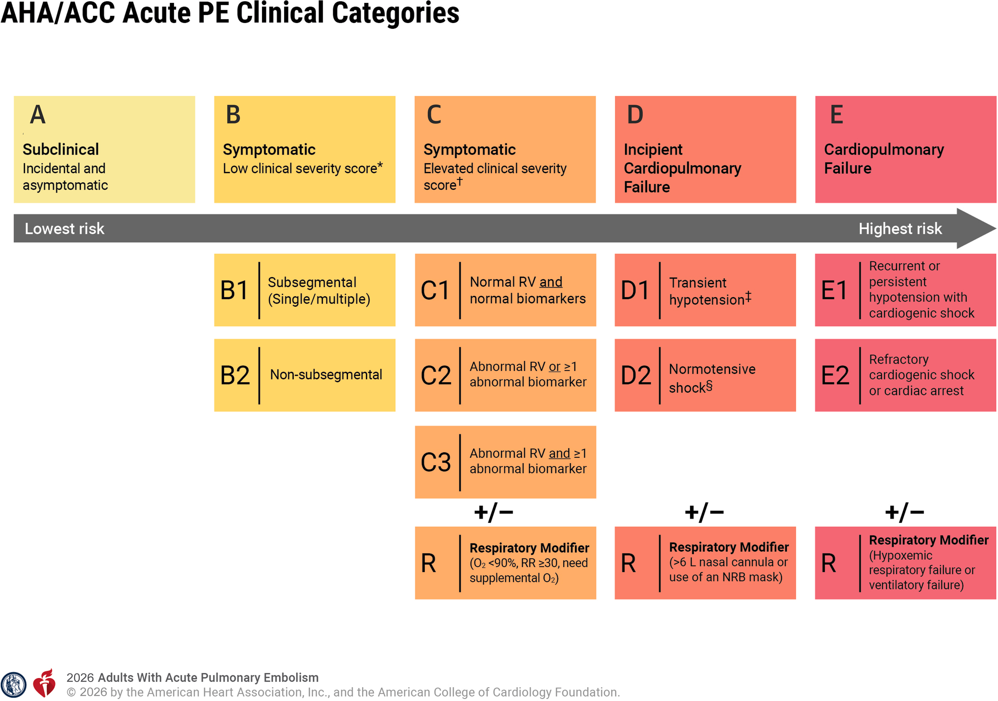
-
Confirmed PE → classify A / B / C / D / E
-
Severity rises from subclinical PE → cardiopulmonary failure
-
Add R respiratory modifier when significant respiratory compromise is present
02 Category A / B — Low Severity
-
Anticoagulation
-
Assess Hestia / PESI / sPESI + clinical suitability for outpatient treatment
-
Category A: Asymptomatic / incidental → generally outpatient
-
Category B: Symptomatic, low clinical severity → early discharge / outpatient treatment when appropriate
03 Category C — Elevated Clinical Severity
-
Hospitalize + anticoagulation
-
PERT assessment
-
C1 → normal RV + normal biomarkers
-
C2 → abnormal RV or ≥1 abnormal cardiac biomarker
-
C3 → abnormal RV and ≥1 abnormal cardiac biomarker
-
No routine systemic thrombolysis
-
Advanced reperfusion → selective / uncertain, especially C3
04 Category D — Incipient Cardiopulmonary Failure
-
D1 → transient hypotension
-
D2 → normotensive shock
-
Anticoagulation + PERT + critical-care monitoring
-
Support RV / systemic perfusion
-
Consider advanced reperfusion: systemic thrombolysis / catheter-directed thrombolysis / mechanical thrombectomy / surgical embolectomy
05 Category E — Cardiopulmonary Failure
-
E1 → persistent / recurrent hypotension + cardiogenic shock
-
E2 → refractory cardiogenic shock / cardiac arrest
-
Immediate resuscitation + advanced reperfusion strategy
-
E1: Systemic thrombolysis / catheter-directed therapy / mechanical thrombectomy / surgical embolectomy
-
E2: Systemic thrombolysis when appropriate ± VA-ECMO / advanced rescue strategy
-
Reference: 2026 AHA/ACC Multisociety PE Guideline — Initial Assessment And Management.
Category A / B — Low Severity
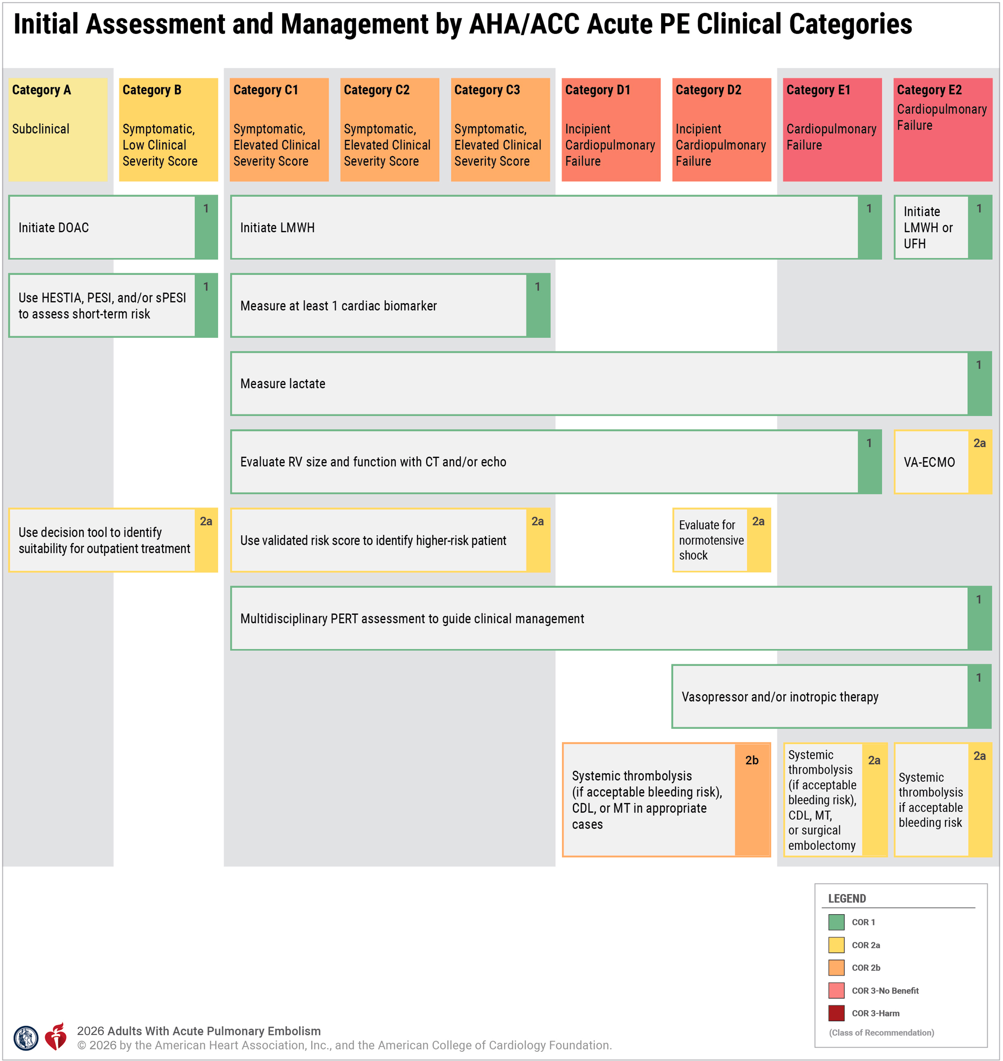
sPESIPrognostic support after PE diagnosis; not a discharge rule
Treatment
-
Anticoagulation: Foundation of treatment unless an absolute contraindication is present
-
DOAC eligible: Direct oral anticoagulant preferred over warfarin
-
Apixaban / rivaroxaban: Can begin treatment without a preceding heparin course; use the acute-VTE regimen
-
Dabigatran / edoxaban: Require an initial parenteral-anticoagulation phase before oral therapy
-
Parenteral therapy required: LMWH preferred over UFH in most eligible patients
-
Special circumstances: Renal / hepatic dysfunction, pregnancy, antiphospholipid syndrome, bleeding risk, drug interactions, and anticipated procedures may change agent selection
-
Reference: 2026 Guideline — Anticoagulation Therapy.
Subsegmental PE — B1
-
B1 distinction matters: The guideline separates subsegmental from more proximal PE because the decision to anticoagulate may differ according to clot location, concomitant DVT, recurrence risk, cardiopulmonary reserve, and bleeding risk
-
Do not assume: Every isolated subsegmental PE automatically receives the same treatment as a proximal PE
-
Reference: 2026 Guideline — Acute PE Clinical Categories.
Outpatient Treatment
-
Category A: Hospitalization is generally unnecessary when no other indication for admission exists
-
Category B: Early discharge / outpatient treatment is generally appropriate when short-term risk is low
-
Risk assessment: Use Hestia, PESI, and/or sPESI plus clinical judgment
-
Safe discharge: Requires immediate access to anticoagulation and rapid, reliable follow-up
-
Admission instead: Significant bleeding risk, oxygen / symptom requirements, important comorbidity, inability to obtain or take anticoagulation, or unreliable follow-up
-
Reference: 2026 Guideline — Suitability For Outpatient Management.
Duration Of Anticoagulation
-
Initial treatment phase: Anticoagulation generally continues for 3–6 months
-
Major reversible provoking factor: Usually stop after the initial treatment phase once the provoking factor has resolved
-
No major reversible risk factor / persistent risk factor: Extended anticoagulation is generally recommended when bleeding risk is acceptable
-
Extended therapy: Reassess recurrence versus bleeding risk periodically rather than assigning an automatic lifetime duration
-
Reference: 2026 Guideline — Anticoagulation By Recurrence Risk.
Category C — Elevated Clinical Severity
Initial Treatment
-
Hospitalize
-
Anticoagulation: Same foundation as above; LMWH preferred when initial parenteral therapy is required
-
PERT: Multidisciplinary PERT assessment recommended for Categories C–E
-
Cardiac biomarker: Obtain troponin and/or BNP to distinguish C1 / C2 / C3
-
RV assessment: CT and/or echocardiography defines RV enlargement / dysfunction
-
Lactate: Helps identify occult hypoperfusion and movement toward Category D
-
Reassess: Worsening BP, perfusion, oxygen requirement, renal function, mental status, or lactate may represent progression toward D
-
Reference: 2026 Guideline — Risk Stratification / PERT.
C1 — Normal RV + Normal Biomarkers
-
Anticoagulation + hospitalization
-
Advanced reperfusion: Not indicated routinely
-
Systemic thrombolysis: Should not be used over anticoagulation alone
C2 — Abnormal RV OR Abnormal Biomarker
-
Anticoagulation + monitored hospitalization
-
Systemic thrombolysis: Should not be used routinely because bleeding harm exceeds demonstrated benefit
-
Catheter-directed thrombolysis / mechanical thrombectomy: Benefit over anticoagulation alone remains unclear in an otherwise stable patient
-
Clinical trajectory: Escalate assessment if perfusion or respiratory status worsens
-
Reference: 2026 Guideline — Advanced Management.
C3 — Abnormal RV + Abnormal Biomarker
-
Anticoagulation + close monitoring
-
PERT: Early involvement because this is the highest-severity Category C subgroup
-
Systemic thrombolysis: Routine use remains unsupported; benefit over anticoagulation alone is uncertain
-
Catheter-directed thrombolysis / mechanical thrombectomy: May be considered selectively, but guideline-level benefit over anticoagulation alone remains uncertain
-
Deterioration: Development of transient hypotension, hypoperfusion, or worsening respiratory failure moves treatment toward the Category D pathway
-
Reference: 2026 Guideline — Systemic And Catheter-Based Reperfusion.
Journal Club — PEITHO — Why We Do Not Routinely Lyse C3-Type Patients
-
Population: Normotensive PE with RV dysfunction + positive troponin
-
Benefit: Tenecteplase decreased death / hemodynamic decompensation at 7 days from 5.6% → 2.6%
-
Harm: Major extracranial bleeding increased, including increased hemorrhagic stroke
-
Bottom line: Systemic thrombolysis decreases deterioration but has not established a mortality benefit sufficient to justify routine treatment of hemodynamically stable patients
-
Reference: PEITHO — Original Randomized Trial.
Journal Club — HI-PEITHO — New Catheter-Directed Evidence
-
Population: Selected intermediate-risk patients with RV dysfunction, myocardial injury, and additional cardiorespiratory distress
-
Treatment: Ultrasound-facilitated catheter-directed fibrinolysis + anticoagulation versus anticoagulation alone
-
Outcome: Seven-day PE-related death / cardiopulmonary decompensation or collapse / recurrent PE 4.0% vs 10.3%
-
Interpretation: Benefit was driven primarily by prevention of clinical deterioration; mortality benefit has not been established
-
Safety: Trial was not powered to firmly exclude a clinically important increase in major bleeding
-
Application: Supports selective catheter-directed therapy in carefully chosen higher-severity patients—not routine intervention for every C2 / C3 PE
-
References: HI-PEITHO — ACC Summary · HI-PEITHO — AHA Data Summary.
Category D — Incipient Cardiopulmonary Failure
Recognize The Transition
-
D1 — Transient hypotension: SBP <90 mm Hg or fall >40 mm Hg that is brief or responds to fluid
-
D2 — Normotensive shock: Evidence of inadequate perfusion despite the absence of persistent hypotension
-
Lactate >2 mmol/L
-
Acute kidney injury / declining urine output
-
Mental-status change
-
Low cardiac index / low MAP or other objective evidence of shock
-
-
R modifier: Escalating oxygen support such as >6 L nasal cannula or nonrebreather use indicates important respiratory compromise
-
Reference: 2026 Guideline — Category D / Hemodynamic Assessment.
Treatment
-
Anticoagulation: Continue unless contraindicated or temporarily altered by the reperfusion strategy
-
PERT / critical care: Immediate multidisciplinary assessment and high-acuity monitoring
-
Vasopressor / inotrope: Use for cardiogenic shock / inadequate systemic perfusion
-
Volume: Consider a limited fluid challenge only when reduced preload is suspected; reassess rather than giving indiscriminate large-volume fluid
-
Advanced reperfusion: Systemic thrombolysis, catheter-directed thrombolysis, mechanical thrombectomy, or surgical embolectomy may be considered
-
Choice of therapy: Balance speed, bleeding risk, anatomy, available expertise, and trajectory
-
Reference: 2026 Guideline — Hemodynamic Pharmacotherapy / Advanced Therapies.
Airway / Respiratory Support
-
Oxygen: Correct clinically significant hypoxemia
-
HFNC: Useful when moderate-severe hypoxemia requires escalating oxygen support
-
Intubation: Avoid unless clinically necessary; induction and positive-pressure ventilation can precipitate RV collapse
-
Prepare before intubation: Vasopressors / inotropes and advanced rescue support should be immediately available
-
Reference: 2026 Guideline — Sedation And Ventilatory Strategies.
Category E — Cardiopulmonary Failure
E1 — Persistent / Recurrent Hypotension With Cardiogenic Shock
-
Resuscitation + PERT / critical care immediately
-
Anticoagulation: Parenteral anticoagulation as appropriate for the planned intervention
-
Systemic thrombolysis: Reasonable when bleeding risk is acceptable
-
Catheter-directed thrombolysis: Reasonable
-
Mechanical thrombectomy: Reasonable
-
Surgical embolectomy: Reasonable
-
Treatment choice: Do not delay a lifesaving available therapy solely to transfer for a theoretically preferred modality
-
Reference: 2026 Guideline — Advanced Therapies.
E2 — Refractory Cardiogenic Shock / Cardiac Arrest
-
Immediate resuscitation
-
Systemic thrombolysis: Reasonable when PE is confirmed / strongly implicated and bleeding risk does not preclude treatment
-
VA-ECMO: Reasonable in refractory cardiogenic shock from known or suspected PE when resources are available
-
Definitive reperfusion after ECMO: Individualize; benefit of routinely adding another advanced therapy after VA-ECMO is not firmly established
-
Cardiac arrest: Treat PE as the reversible cause while continuing high-quality resuscitation
-
Reference: 2026 Guideline — Category E2 / Mechanical Circulatory Support.
Treatment Principles Across Categories
IVC Filter
-
Therapeutically anticoagulated: Do not routinely place an IVC filter
-
Cannot receive anticoagulation: Retrievable IVC filter may be useful when protection from recurrent PE is needed
-
Remove: Retrieve as soon as PE risk has fallen and anticoagulation can be tolerated
-
Reference: 2026 Guideline — IVC Filters.
Reassessment
-
PE category can change: Repeat assessment after treatment and whenever BP, perfusion, oxygen requirement, mental status, renal function, or symptoms worsen
-
C → D → E: Escalation in physiology—not clot size alone—drives escalation in therapy
-
Reference: 2026 AHA/ACC Acute PE Clinical Categories.
In The Pit...
-
A / B → anticoagulate and determine whether the patient can safely go home
-
C → anticoagulate + admit + PERT; RV strain / troponin alone does not mean automatic thrombolysis
-
D → the patient is beginning to fail → support RV / perfusion and actively consider reperfusion
-
E → the patient has cardiopulmonary failure → resuscitate and move immediately toward advanced reperfusion / rescue
-
Watch the trajectory: A patient with a normal systolic BP can still be in shock and belong in Category D
-
Reference: 2026 AHA/ACC Multisociety PE Guideline.
Must-Read References
View Full References
Treatment
-
2026 AHA/ACC Multisociety PE Guideline: Primary current U.S. guideline; introduces the A–E categories and category-directed treatment
-
AHA/ACC Acute PE Clinical Categories: Quick visual reference for A–E, subcategories, and respiratory modifiers
-
PEITHO, 2014: Defines the systemic-thrombolysis benefit / bleeding tradeoff in hemodynamically stable PE with RV injury
-
HI-PEITHO, 2026 — AHA Data Summary: New randomized evidence for catheter-directed fibrinolysis in selected higher-severity intermediate-risk PE
Diagnosis
-
2026 AHA/ACC Multisociety PE Guideline: Probability-based diagnostic framework; Figure 1 connects clinical assessment, PERC, D-dimer, and imaging
-
Courtney et al., 2010: History and examination findings that change PE probability
-
PROPER, 2018: Randomized evaluation of PERC in low-probability ED patients
-
YEARS, 2017: Prospective evaluation of probability-adjusted D-dimer thresholds
-
ADJUST-PE, 2014: Prospective validation of age-adjusted D-dimer
-
Freund et al., 2021: Randomized evaluation of an integrated PERC / YEARS / age-adjusted D-dimer strategy