Gastrointestinal · Clinical algorithm

Abdominal Pain: Upper

Back to contents

Use the zoom controls to enlarge the algorithm, or open the full-screen view.

General Approach

1 Resuscitation / R/O Critical Diagnosis

  • See Abdominal Pain — General for the initial clinical approach to the undifferentiated abdominal-pain patient.
  • Is the patient crashing / unstable? Begin simultaneous resuscitation and evaluation — ABCs, IV/O2/monitor, fluids or blood as indicated, and focused bedside assessment for the cause.
  • Peritoneal signs / concern for surgical abdomen? Consider perforation, ischemia, obstruction, hemorrhage, or other surgical pathology. If stable → focused US / CT based on suspicion. If unstable → urgent surgical evaluation; do not delay definitive care for imaging.
  • Once immediate threats are addressed, move from “Could this patient die from this?” → “Where is the pain coming from?”

2 Emergent Biliary Causes

  • Once the patient is stable and immediate catastrophes are excluded, narrow the upper abdominal source using the history, exam, labs, and focused imaging.
  • Biliary disease → think anatomically: where is the stone?
    • Gallbladder → symptomatic cholelithiasis / biliary colic
    • Cystic duct / gallbladder neck → cholecystitis
    • CBD → choledocholithiasis ± ascending cholangitis
    • Ampulla / transient stone passage → gallstone pancreatitis
  • Historical red flags and labs help localize the process — episodic vs persistent pain, fever, jaundice, Murphy sign, ↑bilirubin / alkaline phosphatase, ↑lipase. RUQ US is usually the first imaging test, then HIDA, MRCP/EUS, or ERCP depending on the suspected disease.

3 Gastric / Pancreatic Causes

  • Acute pancreatitis → epigastric pain radiating to the back + N/V + ↑lipase; then determine etiology, especially gallstones vs alcohol.
  • Peptic ulcer disease → think NSAIDs / H. pylori and, more importantly, whether the ulcer is complicated by bleeding, perforation, or obstruction.
  • If dangerous disease and complicated PUD are excluded, dyspepsia / GERD become more likely outpatient diagnoses.

4 Other Causes / Mimics

  • Do not anchor on the location of tenderness. Always look above and below the area being evaluated.
  • Above the abdomen: ACS may present with epigastric discomfort; lower-lobe pneumonia and PE may present as upper abdominal / lower chest pain.
  • Hepatic / splenic: hepatitis, liver abscess, splenic infarct / injury / rupture may mimic more common upper-GI disease.
  • Below the abdomen: early appendicitis may begin with epigastric / periumbilical pain before migrating to the RLQ.
  • If the GI workup does not fit the clinical picture, re-open the differential rather than defaulting to “gastritis.”

01Resuscitation / R/O Critical Diagnosis

  • See Abdominal Pain — General for the initial approach to the undifferentiated abdominal-pain patient.
  • Is the patient crashing / unstable? Begin resuscitation and diagnostic evaluation simultaneously — ABCs, IV/O2/monitor, fluids or blood as indicated, and rapid bedside assessment for the cause.
  • R/O immediately life-threatening causes — perforation, mesenteric ischemia, hemorrhage/AAA, bowel obstruction, ectopic pregnancy when applicable, or other surgical catastrophe.
  • Peritoneal signs / concern for surgical abdomen?
    • Stable → focused US / CT based on the suspected diagnosis.
    • Unstable → urgent surgical evaluation; do not delay definitive care for imaging.
  • Reassess after initial stabilization. Once the immediate threats are addressed, shift from “Could this patient die from this?” → “Where is the pain coming from?”
  • Then use the location, pain pattern, exam, and focused labs/imaging to narrow toward biliary, pancreatic, gastric, or other causes.

02Emergent Biliary Causes

Biliary Tract Disease

Cholelithiasis

  • Gallstones are common and usually asymptomatic.
  • Most gallstones are incidental and never cause symptoms. Merck
  • Finding gallstones does not automatically mean they are causing the patient's abdominal pain.
  • The clinical pain pattern must fit biliary disease before attributing symptoms to cholelithiasis.

Biliary Colic

  • Symptomatic cholelithiasis → transient cystic-duct obstruction Merck
Red Flags — What Moves The Needle?
  • Episodic RUQ or epigastric pain
    • Pain is typically steady during the episode, despite the term “colic.”
    • Resolves spontaneously over hours.
  • May be postprandial.
    • Fatty foods are not a specific diagnostic clue.
  • Patient is generally well appearing and afebrile.
  • May have nausea and mild RUQ tenderness.
  • Labs are usually normal in uncomplicated biliary colic.
    • Abnormal inflammatory, liver, or pancreatic studies should raise concern for a complication.
Diagnosis
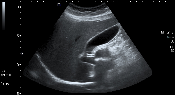
  • RUQ ultrasound ACR
  • Typical biliary pain + gallstones without inflammatory or obstructive findings supports uncomplicated symptomatic cholelithiasis / biliary colic.
Treatment / Disposition
  • Pain control
  • Outpatient surgical referral for symptomatic gallstones. SAGES

Cholecystitis

  • Persistent cystic-duct obstruction → gallbladder inflammation
Red Flags — What Moves The Needle?
  • Persistent RUQ or epigastric pain, typically >6 hours
  • RUQ tenderness / Murphy sign
    • A positive Murphy sign supports the diagnosis but is not diagnostic by itself.
  • Nausea / vomiting ± fever
  • Leukocytosis
  • LFTs may be normal or mildly elevated.
    • Marked bilirubin / alkaline phosphatase elevation → consider CBD obstruction / choledocholithiasis
Diagnosis
  • RUQ ultrasound ACR
    • Gallstones
    • Gallbladder wall thickening
    • Pericholecystic fluid
    • Sonographic Murphy sign
  • If ultrasound is equivocal but clinical suspicion remains → consider HIDA
Treatment / Disposition
  • Supportive care + antibiotics
  • Surgery consultation → early laparoscopic cholecystectomy during the index admission WSES 2020
Pitfall
  • Critically ill patient + unexplained sepsis / RUQ findings → consider acalculous cholecystitis even without gallstones

Choledocholithiasis

  • Gallstone in the common bile duct → partial or complete biliary obstruction Merck
Red Flags — What Moves The Needle?
  • Jaundice
  • Biliary-type RUQ / epigastric pain
  • Elevated bilirubin / alkaline phosphatase ± jaundice Merck
  • Fever / systemic illness → think ascending cholangitis, not uncomplicated choledocholithiasis.
Diagnosis
  • RUQ ultrasound
    • CBD dilation?
    • CBD stone seen?
    • Ultrasound may suggest obstruction without directly visualizing the stone.
  • Persistent suspicion with uncertain CBD stone → MRCP or EUS rather than diagnostic ERCP. ASGE
  • Strong evidence / high probability of retained CBD stone → ERCP for diagnosis and duct clearance. ASGE
Treatment / Disposition
  • GI consultation
  • ERCP / duct clearance when indicated

Gallstone Pancreatitis

  • Gallstone migration through the ampulla → acute pancreatitis
  • See Acute Pancreatitis
  • RUQ ultrasound to evaluate for gallstones / biliary etiology.
  • Evaluate for persistent CBD obstruction or ascending cholangitis.
    • If the retained CBD stone is uncertain → MRCP or EUS
    • ERCP when persistent obstruction or cholangitis is present
  • Do not routinely perform urgent ERCP for gallstone pancreatitis alone when there is no cholangitis or persistent biliary obstruction. ASGE

Ascending Cholangitis

  • Biliary obstruction + infection → cholangitis → potentially sepsis / septic shock
Red Flags — What Moves The Needle?
  • Charcot triad: RUQ pain + fever + jaundice
    • Strongly supports the diagnosis, but absence of the complete triad does not exclude cholangitis. Tokyo Guidelines
  • Reynolds pentad: Charcot triad + hypotension + altered mental status
    • Think severe / septic cholangitis.
  • More broadly, think cholangitis when there is:
    • Systemic inflammation
    • Cholestasis
    • Evidence of biliary obstruction Tokyo Guidelines
Diagnosis
  • RUQ ultrasound → biliary dilation / obstructing stone?
  • MRCP / EUS may further define obstruction when needed.
  • Do not wait for the full Charcot triad if the overall picture suggests an infected obstructed biliary system.
Treatment / Disposition
  • Resuscitation + broad-spectrum antibiotics
  • Urgent GI consultation → biliary drainage, usually ERCP ASGE
  • Endoscopic drainage is preferred when feasible; percutaneous drainage is an alternative when ERCP cannot be performed.

03Gastric / Pancreatic Causes

Acute Pancreatitis

Red Flags — What Moves The Needle?

  • Epigastric pain, usually constant, often radiating to the back
  • Nausea / vomiting
  • Epigastric tenderness ± guarding
  • Look for the etiology:
    • Gallstones / biliary disease
    • Alcohol
    • If neither explains the episode → check triglycerides ACG 2024
Board Pearls
  • Cullen sign → periumbilical ecchymosis
  • Grey Turner sign → flank ecchymosis
  • Rare findings that suggest severe intra-abdominal / retroperitoneal hemorrhagic disease; memorable but not sensitive diagnostic findings. Review

Diagnosis

  1. Characteristic abdominal pain
  2. Lipase or amylase ≥3× upper limit of normal
  3. Characteristic imaging
  • Lipase is the preferred pancreatic enzyme.
    • The degree of lipase elevation does not determine disease severity.
  • Do not routinely CT a straightforward case.
    • CT → diagnosis unclear or failure to improve clinically within 48–72 hours ACG 2024
  • RUQ ultrasound to evaluate for gallstone / biliary etiology.

Severity — Revised Atlanta Classification

  • Local complications include peripancreatic fluid collections, pancreatic/peripancreatic necrosis, pseudocyst, and walled-off necrosis. Revised Atlanta Classification
  • Organ failure / persistent SIRS → monitored or ICU-level setting as appropriate ACG 2024

Special Etiology — Hypertriglyceridemia

  • If gallstones and significant alcohol use are absent, check serum triglycerides.
  • TG >1,000 mg/dL strongly supports hypertriglyceridemia as the cause. ACG 2024
  • Treat the pancreatitis supportively and correct the metabolic cause.
  • Uncontrolled diabetes / significant hyperglycemia → insulin infusion is appropriate and will also lower triglycerides.
  • In patients without diabetes, routine insulin infusion is not established as standard therapy. Endocrine Society
  • Plasmapheresis is not routine first-line therapy; consider only selected extraordinary or refractory cases. Endocrine Society
  • After the acute episode, address secondary causes and institute long-term triglyceride-lowering therapy, commonly including a fibrate when appropriate.

Treatment / Disposition

  • Analgesia
  • Moderate, reassessment-driven IV fluids; LR preferred ACG 2024
    • Avoid indiscriminate large-volume resuscitation.
    • The WATERFALL trial found substantially more fluid overload with aggressive hydration without improved clinical outcomes. WATERFALL
  • Early oral feeding as tolerated rather than routine prolonged NPO / bowel rest. ACG 2024
  • No prophylactic antibiotics for sterile pancreatic necrosis. ACG 2024
  • Necrosis alone does not mandate early surgery.
    • If intervention is required and the patient is stable, favor delayed, minimally invasive / step-up management rather than early open surgery. ACG 2024
  • Gallstone pancreatitis → see Biliary Tract Disease
  • Pancreatic Fluid Collections
    • Intervention is driven primarily by symptoms, infection, obstruction, bleeding, or other complications, not size alone. ASGE

Dyspepsia / GERD / Peptic Ulcer Disease

  • Upper abdominal burning, epigastric discomfort, and reflux symptoms overlap.
  • After excluding important alternate diagnoses, consider PUD, dyspepsia, and GERD.

Peptic Ulcer Disease

  • Think PUD → H. pylori or NSAIDs
Red Flags — What Moves The Needle?
  • Burning / gnawing epigastric pain
  • NSAID / aspirin use
  • Prior PUD / H. pylori (major causes of PUD. Merck)
  • Meal association may occur, but “gastric worse with food / duodenal better with food” is not reliable enough to diagnose PUD
  • Hematemesis / melena / anemia / hemodynamic change → GI bleeding
  • Sudden severe pain / peritoneal findings → perforation
  • Persistent vomiting / early satiety / PO intolerance → gastric outlet obstruction
Diagnosis
  • In ED suspected PUD based on symptoms / risk factors after dangerous alternatives have been excluded.
  • H. pylori: patients with current or prior PUD outpatient testing and treatment
  • PUD confirmed by EGD
Treatment / Disposition
For Uncomplicated Suspected PUD
  • PPI (Omeprazole 20 mg PO daily or pantoprazole 40 mg PO daily)
  • Stop nonessential NSAIDs
  • Arrange H. pylori testing / outpatient follow-up
  • Persistent or concerning symptoms → GI / EGD
Complicated PUD
  • Bleeding → See Upper GI Bleeding
  • Perforation → See Intestinal Perforation
  • Gastric outlet obstruction → admission / GI ± surgery

Dyspepsia / GERD

Dyspepsia
  • Epigastric pain / burning
  • Postprandial fullness
  • Early satiety
  • ± nausea
Treatment
  • <60 years → consider outpatient H. pylori test-and-treat
    • If negative or symptoms persist → PPI trial
  • New dyspepsia ≥60 → GI / upper endoscopy ACG/CAG
GERD
  • Heartburn
  • Regurgitation / water brash
  • Often postprandial or positional
  • May worsen lying down
  • Chest pain → exclude cardiac disease before attributing it to GERD
Treatment
  • For classic GERD without alarm features:
    • PPI once daily before a meal × 8 weeks ACG
  • Antacids / H2 blockers are reasonable for mild intermittent symptoms; PPI is preferred for persistent GERD or suspected ulcer disease.
Consider GI / EGD For
  • GI bleeding / anemia
  • Dysphagia
  • Unexplained weight loss
  • Persistent vomiting
  • Persistent / recurrent symptoms despite treatment
In The Pit...
  • Uncomplicated PUD / dyspepsia / GERD → PPI + stop nonessential NSAIDs + outpatient H. pylori / GI follow-up as appropriate
  • Bleeding → Upper GI Bleeding
    Perforation → Intestinal Perforation
    Obstruction → admission / GI ± surgery

04Other Causes / Mimics

  • Consider disease above / below area of tenderness

Cardiopulmonary

  • ACS → epigastric / upper abdominal discomfort may be anginal equivalent AHA/ACC
  • Lower-lobe pneumonia → fever / cough / dyspnea / hypoxia ± upper abdominal pain Merck
  • PE → pleuritic upper abdominal / lower chest pain + dyspnea / hypoxia / VTE risk

Hepatic / Splenic

  • Acute hepatitis → RUQ pain + ↑AST/ALT ± jaundice Merck
  • Liver abscess → RUQ pain + fever / sepsis
  • Splenic infarct / injury / rupture → LUQ pain ± left shoulder pain; trauma / embolic risk Merck

GI

Cases

Pop Quiz Hotshot

Must-Read References

View Full References

Additional Cited Guidance

Privacy & analytics choices