General Approach
1 Resuscitation / R/O Critical Diagnosis
- See Abdominal Pain — General for the initial clinical approach to the undifferentiated abdominal-pain patient.
- Is the patient crashing / unstable? Begin simultaneous resuscitation and evaluation — ABCs, IV/O2/monitor, fluids or blood as indicated, and focused bedside assessment for the cause.
- Peritoneal signs / concern for surgical abdomen? Consider perforation, ischemia, obstruction, hemorrhage, or other surgical pathology. If stable → focused US / CT based on suspicion. If unstable → urgent surgical evaluation; do not delay definitive care for imaging.
- Once immediate threats are addressed, move from “Could this patient die from this?” → “Where is the pain coming from?”
2 R/O Emergent Causes
- Once the patient is stable and the critical abdominal diagnoses are addressed → localize the pain and narrow the source.
- RLQ pain / tenderness → R/O appendicitis. Migration of pain, focal RLQ tenderness, peritoneal irritation, and an inflammatory response increase suspicion.
- LLQ pain / tenderness → R/O diverticulitis. Focal LLQ findings ± fever / inflammatory markers suggest the diagnosis; CT A/P confirms diverticulitis and determines uncomplicated vs complicated disease.
- Do not stop with GI pathology. Lower abdominal pain frequently overlaps with GU and pelvic disease.
- GU → nephrolithiasis, pyelonephritis / cystitis; male lower abdominal or groin pain → consider acute scrotum / testicular torsion.
- GYN → ectopic pregnancy, ovarian torsion, PID / TOA, ovarian cyst. Pregnancy test early → pelvic US based on suspicion.
3 Other GI Causes
- If appendicitis, diverticulitis, GU, and GYN disease do not explain the presentation → broaden to other inflammatory, infectious, functional, and focal GI causes.
- Known IBD + abdominal pain → do not anchor on the IBD. First ask: other diagnosis vs active flare vs IBD complication?
- If flare → determine severity.
- Severe / focal pain, vomiting / distention, sepsis, or peritoneal findings → R/O obstruction, abscess, perforation, toxic megacolon → CT.
- Colitis → infectious vs inflammatory vs ischemic. Diarrhea / bloody stool and the clinical context help determine which pathway to pursue.
- Common recurrent / lower-risk GI syndromes include gastroenteritis, constipation, IBS, and cannabinoid hyperemesis syndrome. These become more likely when the clinical pattern fits and dangerous structural disease has been excluded.
4 Appendicitis / Diverticulitis Mimics
- Focal GI mimics include mesenteric adenitis, typhlitis, intussusception, epiploic appendagitis, omental infarction, and hernia. Think of these when focal pain resembles appendicitis / diverticulitis but the overall picture or imaging points elsewhere.
01Resuscitation / R/O Critical Diagnosis
- See Abdominal Pain — General for the initial approach to the undifferentiated abdominal-pain patient.
- Is the patient crashing / unstable? Begin resuscitation and diagnostic evaluation simultaneously — ABCs, IV/O2/monitor, fluids or blood as indicated, and rapid bedside assessment for the cause.
- R/O immediately life-threatening causes — perforation, mesenteric ischemia, hemorrhage/AAA, bowel obstruction, ectopic pregnancy when applicable, or other surgical catastrophe.
- Peritoneal signs / concern for surgical abdomen?
- Stable → focused US / CT based on the suspected diagnosis.
- Unstable → urgent surgical evaluation; do not delay definitive care for imaging.
- Reassess after initial stabilization. Once the immediate threats are addressed, shift from “Could this patient die from this?” → “Where is the pain coming from?”
- Then use the location, pain pattern, exam, and focused labs/imaging to narrow toward appendiceal, diverticular, GU/GYN, or other GI causes.
02R/O Emergent Causes
Acute Appendicitis
Clinical Presentation
- Periumbilical / poorly localized pain → migrates to RLQ → constant / sharper
- RLQ tenderness / localized peritoneal irritation
- McBurney point tenderness
- Guarding / rebound
- Anorexia, N/V ± constipation / diarrhea
- Fever / tachycardia ± ↑WBC / CRP
- Rovsing sign → LLQ palpation produces RLQ pain
- Psoas sign → pain with hip extension / flexion; may suggest retrocecal appendix
- Obturator sign → pain with flexion / internal rotation of hip
- Rovsing / psoas / obturator → supportive findings; absence does not exclude appendicitis
Red Flags — What Moves The Needle?
- Migration of pain → RLQ
- Focal RLQ tenderness / peritoneal irritation
- Anorexia / N/V
- Inflammatory response → fever, ↑WBC / CRP
- Combination of migration + localized peritoneal findings + inflammatory response → ↑ suspicion
- Extremes of age / pregnancy / immunocompromised → may have atypical or subtle presentation
Diagnosis
Imaging Based On Patient Population
Nonpregnant adult → CT A/P, usually with IV contrast
IDSA 2024 — Adult Appendicitis ImagingChild / adolescent → US first
- Equivocal / nondiagnostic US + persistent suspicion → MRI or CT
- Nonvisualized / partially visualized appendix = equivocal, not negative
ACEP 2023 — Appendicitis Clinical Policy
Pregnant → US first ± MRI
- Equivocal US + persistent suspicion → MRI without contrast
ACR — RLQ Pain / Pregnancy Imaging
- Equivocal US + persistent suspicion → MRI without contrast
Clinical scores → may help risk-stratify; do not use alone to exclude appendicitis / avoid imaging.
Treatment / Disposition
- Analgesia
- Antibiotics
- Surgery consultation
- Laparoscopic appendectomy → standard definitive treatment
- Complicated appendicitis — perforation / diffuse peritonitis / abscess / phlegmon / sepsis → source-control pathway
Nonoperative Treatment
- Selected uncomplicated appendicitis → antibiotics alone may be considered with surgery involvement + shared decision-making.
Journal Club — CODA Trial
- Antibiotics noninferior to appendectomy for 30-day health status
- 29% → appendectomy by 90 days
- Appendicolith:
- 41% appendectomy by 90 days vs 25% without appendicolith
- ↑ complications with antibiotics-first treatment
CODA Trial — Antibiotics vs Appendectomy
- Appendicolith → higher risk of antibiotics-first failure / complications
- Nonoperative treatment applies to selected uncomplicated disease; complicated appendicitis requires a separate source-control strategy.
In The Pit...
- RLQ pain / tenderness +
- Migration / peritoneal findings / inflammatory response → Image
- Adult → CT
- Child → US → MRI / CT if equivocal
- Pregnant → US → MRI if equivocal
- Nondiagnostic US ≠ negative study
- Confirmed appendicitis → surgery consultation
- Can't Miss Diagnosis: Any right lower quadrant abdominal pain or tenderness needs a workup for appendicitis
Diverticulitis
Clinical Presentation
- Acute / subacute LLQ pain + focal tenderness
- Fever ± ↑WBC / CRP
- Change in bowel habits ± nausea
- Clinical findings supportive but nonspecific — H&P alone accurately predicts diverticulitis in only ~40–65%
AGA — Clinical Presentation
Red Flags — What Moves The Needle?
- Focal LLQ pain / tenderness + inflammatory response
- Peritoneal signs / severe pain → perforation?
- Fever / sepsis / toxic appearance → complicated disease
- Persistent vomiting / PO intolerance
- Immunocompromised / frail / significant comorbidity
- Failure to improve → abscess / other complication?
- Blood in stool / anemia / weight loss → alarm feature / alternate diagnosis
Diagnosis
- CT A/P → confirm diagnosis + uncomplicated vs complicated
- IV contrast usually appropriate; helps define complications
IDSA 2024 — Diverticulitis Imaging
- IV contrast usually appropriate; helps define complications
- First episode / uncertain diagnosis / severe presentation / failure to improve → CT
- Known recurrent, classic mild disease → repeat imaging not always necessary
AGA — CT Indications - CT unavailable / contraindicated → US or MRI
- Pregnant → US or MRI
IDSA 2024 — Pregnancy
Uncomplicated Vs Complicated
- Uncomplicated → localized diverticular inflammation
- Complicated → abscess / phlegmon / perforation / fistula / obstruction ± systemic illness
ACP 2022
Treatment / Disposition
- Uncomplicated / immunocompetent / mild
- Outpatient treatment if well appearing, stable, tolerating PO, pain controlled + reliable follow-up
- Antibiotics selective — not routine
- AGA: selective antibiotics in immunocompetent mild uncomplicated disease
AGA — Antibiotic Recommendation - ACP: selected uncomplicated patients → initial management without antibiotics
ACP 2022 — No-Antibiotic Option
- AGA: selective antibiotics in immunocompetent mild uncomplicated disease
- Journal Club — Au & Aly 2019 meta-analysis: 9 studies / 2,505 patients → no significant difference in additional treatment, readmission, surgery / radiologic intervention, recurrence, or complications without antibiotics
DOI | PubMed - Clear liquids for comfort → advance as tolerated
AGA — Diet
- Antibiotics / admission more likely
- Complicated disease
- Sepsis / systemic illness
- Immunocompromised / frail / significant comorbidity
- Refractory symptoms / persistent vomiting
- Unable to tolerate PO
- Failed outpatient treatment
- If antibiotics → gram-negative + anaerobic coverage
AGA — Higher-Risk Disease
Complications
- Abscess → antibiotics ± drainage
- Perforation / generalized peritonitis → surgery / source control
- Fistula
- Obstruction
Follow-Up
- Colonoscopy after complicated diverticulitis and generally after first uncomplicated episode, depending on prior colonoscopy / disease course
- Usually delay 6–8 weeks after resolution
AGA — Colonoscopy After Diverticulitis
In The Pit...
- LLQ pain / tenderness + inflammatory findings → consider diverticulitis
- CT → confirm + uncomplicated vs complicated
- Uncomplicated / healthy / tolerating PO → outpatient ± no antibiotics
- Complicated / high-risk / septic → antibiotics + admission / source control
- Failure to improve → reassess / image for complication
Genitourinary Causes
- Lower abdominal pain may originate from the urinary tract or scrotum.
Pyelonephritis / cystitis → dysuria, frequency, fever, flank / CVA tenderness
See Low Back Pain AlgorithmNephrolithiasis → colicky flank / lower abdominal pain ± hematuria, N/V
See Low Back Pain AlgorithmTesticular torsion / epididymitis / other acute scrotal pathology → lower abdominal or groin pain may precede or accompany scrotal symptoms
See Acute Scrotum Algorithm
Gynecologic Causes
- In reproductive-age females, always consider a pelvic source of lower abdominal pain.
Ectopic pregnancy
Ovarian torsion
PID / tubo-ovarian abscess
Ovarian / hemorrhagic cyst
Fibroids / other pelvic pathology
- Pregnancy test early → pelvic US based on suspicion
See Pelvic Pain Algorithm — Coming soon
03Other GI Causes
Inflammatory Bowel Disease
- Main ED question → uncomplicated flare vs acute complication?
Clinical Presentation
Crohn disease → abdominal pain ± diarrhea; RLQ pain with terminal ileitis
Ulcerative colitis → abdominal pain + bloody diarrhea / urgency
Fever / tachycardia ± ↑WBC / CRP → active / severe inflammation
New focal / severe pain or systemic illness → look for complication rather than assuming “flare”
Red Flags — What Moves The Needle?
Pain + vomiting / distention → obstruction / stricture?
Fever + focal tenderness / sepsis → abscess?
Peritoneal signs → perforation?
Severe / ongoing hematochezia → hemorrhage?
Bloody diarrhea + systemic toxicity → acute severe UC / toxic megacolon?
Steroids / immunosuppression → may blunt peritoneal findings
Diagnosis
Suspected acute IBD complication → CT A/P with IV contrast
WSES-AAST — Emergency IBD imagingSuspected flare with significant diarrhea → exclude infection / C. difficile
ASUC → test for C. difficile
ACG 2025 UC — Original Guideline
Acute Severe Ulcerative Colitis
- Frequent bloody diarrhea + systemic toxicity → ASUC
Admit + GI
C. difficile testing
Assess for toxic megacolon
IV corticosteroids
No routine broad-spectrum antibiotics unless infection / sepsis / perforation concern
ACG 2025 UC — AntibioticsAvoid NSAIDs / opioids / anticholinergics → may worsen colonic dilation / toxicity
ACG 2025 UC — Medications To Avoid
Toxic Megacolon
- Colonic dilation + systemic toxicity → life-threatening complication of severe inflammatory / infectious colitis
Ill appearing, abdominal distention, fever / tachycardia, bloody diarrhea
ASUC → assess specifically for toxic megacolon
ACG 2025 UC — Toxic MegacolonResuscitation + GI / surgery
Toxic megacolon / perforation / severe refractory hemorrhage / refractory disease → surgical pathway
Crohn Complications
Stricture / obstruction
Abscess
Fistula / penetrating disease
Perforation
Severe hemorrhage
Suspected complication → CT + GI / surgery ± IR
Abscess → antibiotics + drainage / source control as appropriate
ACG 2025 Crohn Disease — Original Guideline
In The Pit...
- Other diagnosis? → flare? → how sick? → complication?
- Known IBD + abdominal pain...
- 1. Is this actually the IBD?
- Do not anchor on IBD
- Consider usual abdominal-pain differential → appendicitis, diverticulitis, GU/GYN, gastroenteritis, dyspepsia, etc.
- 2. Does this fit an IBD flare?
- UC → ↑ stool frequency + hematochezia / urgency / tenesmus
- Crohn → abdominal pain + diarrhea ± weight loss / inflammatory findings
- Suspected flare with diarrhea → exclude infection, especially C. difficile
- 3. If this is IBD, how sick?
- Mild / uncomplicated
- Well appearing, stable, tolerating PO
- No significant bleeding / anemia / sepsis
- No peritoneal signs / obstruction
- Supportive care + outpatient GI follow-up / disease-directed plan
- Moderate / severe
- Significant bloody diarrhea, dehydration / PO intolerance, anemia, fever / tachycardia, systemic toxicity
- Admit + GI
- ASUC
- ≥6 bloody stools/day + systemic toxicity
- Admit + GI
- C. difficile testing
- IV corticosteroids
- Assess for toxic megacolon
- 4. Could there be an IBD complication?
- Focal / severe pain
- Persistent vomiting / distention → obstruction / stricture?
- Fever / sepsis / focal tenderness → abscess?
- Peritoneal signs → perforation?
- Toxic megacolon / major hemorrhage?
- → CT A/P with IV contrast + admit + GI / surgery ± IR
- Antibiotics → infection / abscess / sepsis / perforation concern; not routine uncomplicated flare
Colitis
- Infectious colitis → diarrhea ± fever / blood → See Diarrhea — Coming soon
- Inflammatory colitis → See IBD
- Ischemic colitis → abdominal pain + hematochezia, especially older / vascular-risk patient → consider CT See Mesenteric Ischemia
Other Common GI Causes
- Gastroenteritis → N/V/D + benign abdominal exam → See Diarrhea — Coming soon
- Constipation / fecal loading → abdominal discomfort / distention + ↓ stool output
- IBS → recurrent abdominal pain + altered bowel habits; no alarm features
- Cannabinoid hyperemesis syndrome → chronic cannabis use + recurrent severe N/V ± abdominal pain; consider after excluding important alternate causes
04Appendicitis / Diverticulitis Mimics
- Mesenteric adenitis → RLQ pain, often after viral illness; diagnosis of exclusion
- Typhlitis → RLQ pain + fever in neutropenic / immunocompromised patient → potentially serious
- Intussusception → episodic / colicky abdominal pain ± vomiting; especially pediatrics
- Epiploic appendagitis → focal LLQ or RLQ pain; CT mimic of diverticulitis / appendicitis; usually supportive treatment
- Omental infarction → focal abdominal pain; CT diagnosis; usually supportive treatment
- Hernia → focal pain / mass; incarceration / strangulation → surgical emergency
Cases
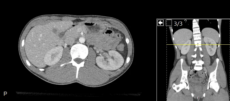
Pop Quiz Hotshot
Must-Read References
View Full References
- IDSA 2024: Intra-Abdominal Infections — Imaging guidance for suspected appendicitis, diverticulitis, and intra-abdominal complications.
- ACEP 2023: Appendicitis Clinical Policy — Emergency-department evaluation, including equivocal pediatric ultrasound and the limitations of clinical scores.
- CODA Trial: Antibiotics Versus Appendectomy — Evidence for shared decision-making about antibiotics-first treatment and the importance of an appendicolith.
- AGA Clinical Practice Update: Medical Management Of Colonic Diverticulitis — Practical guidance on imaging, selective antibiotics, diet, and follow-up colonoscopy.
- Au And Aly 2019: Treatment Of Uncomplicated Acute Diverticulitis Without Antibiotics — Systematic review and meta-analysis supporting selective antibiotic use in appropriate patients.
- ACG 2025: Ulcerative Colitis In Adults — Guidance for acute severe ulcerative colitis and its complications.
- ACG 2025: Management Of Crohn Disease In Adults — Disease management and pathways for important Crohn complications.
Additional Cited Guidance
- ACR Appropriateness Criteria: Right Lower Quadrant Pain — Imaging choices for nonpregnant and pregnant patients.
- ACP 2022: Diverticulitis Guidelines — Diagnosis, outpatient care, and selective treatment without antibiotics.
- WSES-AAST: Management Of Inflammatory Bowel Disease In The Emergency Setting — Assessment and management of acute IBD complications.