THE CHIEF
COMPLAINT
Back to contents

Cardiology · Clinical algorithm

Chest Pain

Use the zoom controls to enlarge the algorithm, or open the full-screen view.

General Approach

01 Initial Evaluation / Assessment

  • Resuscitation, ECG, and evaluation for critical causes proceed together. Do not finish one before starting the others.

Unstable?

  • Shock / hypotension / altered mental status / severe respiratory distress → immediate resuscitation
  • Resus: ABCs, IV access, monitor, pulse oximetry; oxygen for hypoxemia
    • Airway / ventilatory support?
    • Fluids / blood / vasopressors according to the suspected cause; reassess the response
  • Critical clues — identify what is causing the instability
    • ECG / monitor: STEMI / acute coronary occlusion pattern? Arrhythmia?
    • POCUS: Tamponade, LV dysfunction, RV strain, aortic-root / dissection clues, lung sliding / pneumothorax
    • Focused exam: Asymmetric breath sounds, pulse deficits, new murmur, focal neurologic deficit
    • Legs: Unilateral swelling / DVT signs
    • Portable CXR: Pneumothorax, pulmonary edema, pleural fluid, mediastinal abnormality; do not delay an immediate intervention
  • Reference: ACEP RUSH.

ECG — Stable Or Unstable

  • Obtain and interpret within 10 minutes, including patients who appear well
  • STEMI / acute coronary occlusion pattern → activate the cath-lab / reperfusion pathway
    • Do not wait for troponin
  • Persistent / recurrent symptoms + nondiagnostic ECG → repeat ECG
  • Reference: ACC/AHA 2025.

02 R/O Emergent Causes

  • STEMI / acute coronary occlusion
    • Substernal pressure / heaviness ± arm / jaw radiation, dyspnea, diaphoresis, or nausea increases suspicion
    • ECG identifies the reperfusion pathway—not the pain description alone. Activate immediately when indicated; primary PCI preferred.
  • Aortic dissection
    • Abrupt severe chest / back pain, maximal at onset, especially with pulse / BP differential, neurologic deficit, or new AR murmur
    • Meaningful suspicion → CTA aorta
    • Unstable → immediate aortic-team / surgical involvement + bedside echo / TEE; CT only when safe and without delaying definitive care.
  • Pulmonary embolism
    • Pleuritic pain / unexplained dyspnea / hypoxemia, supported by unilateral leg findings or VTE risk factors
    • Low probability → PERC; negative → no further PE testing
    • Low, PERC-positive OR intermediate probability → D-dimer; above the applicable validated cutoff → imaging
    • High probability → CTPA directly
    • Follow the PE diagnostic pathway; instability requires simultaneous resuscitation and urgent evaluation.
  • Pericarditis / myocarditis
    • Pericarditis: Pleuritic / positional pain ± friction rub
    • Myocarditis: Chest pain with new HF, arrhythmia, or syncope; pericarditis need not be present
    • ECG + troponin + CRP / ESR + echo
    • Tamponade with compromise → urgent drainage; myocardial involvement / high-risk findings → cardiology and monitored care.
  • Esophageal perforation
    • Forceful vomiting → sudden chest / upper abdominal pain, or new pain after instrumentation
    • CXR may provide a clue; a normal film does not exclude
    • CT esophagram + NPO / IV antibiotics / early thoracic surgery involvement.
  • Pneumothorax
    • Sudden pleuritic pain / dyspnea + asymmetric breath sounds
    • Stable → lung US / CXR
    • Suspected tension + instability → immediate decompression; do not wait for imaging.

03 R/O ACS

  • ECG / clinical assessment → choose the pathway
    • STEMI / acute coronary occlusion pattern → reperfusion, as above
    • Ischemic ECG / high-risk clinical findings → high-risk suspected NSTE-ACS pathway: Cardiology, monitored care, serial ECGs / hs-cTn
    • Concerning / equivocal ECG → cardiologist review + serial ECGs ± urgent echo
    • Nonischemic ECG + clinically stable → high-sensitivity troponin clinical decision pathway—hs-cTn CDP
  • hs-cTn CDP → determine the next step
    • Use your institution’s assay, sampling times, cutoffs, and delta criteria
    • Rule-out / low risk → discharge with follow-up when the complete assessment supports it
    • Intermediate / observation zone → repeat hs-cTn at 3–6 hours + reassess; additional testing as needed
    • Abnormal / high risk → additional evaluation / usually admission
      • Elevated troponin establishes myocardial injury—not automatically NSTEMI
  • Reference: ACC 2022.

04 Other Causes / Disposition

  • Other cardiac causes: Stable angina or another cardiac condition; a new or worsening ischemic pattern returns the patient to ACS evaluation
  • GI: Reflux / esophageal disease / upper abdominal pathology
  • Chest wall / musculoskeletal: Localized tenderness or movement-related pain; reproducibility alone does not exclude ACS
  • Pulmonary / infectious: Pneumonia or another pulmonary process
  • Anxiety-related symptoms: Assess in context—not simply because the initial tests are negative
  • Before discharge: Reassess symptoms, vital signs, and examination. Persistent concerning findings require further evaluation; a low-risk ACS assessment does not exclude another dangerous diagnosis
  • Follow-up / return precautions: Address recurrent or worsening symptoms and unresolved diagnostic concerns.

01Resuscitation / Initial Evaluation

  • Resuscitation and evaluation proceed together. Obtain the ECG and focused bedside assessment during resuscitation—not afterward.
  • Early ECG applies to stable and unstable patients.

Unstable?

  • Shock / poor perfusion: Hypotension, altered mental status, cool / clammy extremities
  • Severe respiratory compromise
  • Unstable tachyarrhythmia / bradyarrhythmia
  • Acute heart failure / pulmonary edema with clinical compromise
  • Reference: ACC 2022.

Immediate Resuscitation

  • ABCs: Assess and support airway, breathing, and circulation.
  • Establish IV access, cardiac monitoring, and pulse oximetry.
  • Oxygen: Treat hypoxemia; assess the need for airway / ventilatory support.
  • Circulatory support: Give fluids, blood, or vasopressors according to the suspected cause—not automatic large-volume fluids.
    • Reassess perfusion and response; use bedside cardiac findings to guide further support.
  • No pulse: Begin the cardiac arrest pathway.
  • Activate the appropriate specialist team early when an immediately treatable cause is identified.
  • References: ACC/AHA 2025 · ACEP RUSH.

Critical Clues — During Resuscitation

  • ECG / monitor
    • Look for a STEMI / acute coronary occlusion pattern.
    • Identify an arrhythmia causing instability.
  • POCUS — Heart / Lungs
    • Assess for pericardial effusion with tamponade physiology.
    • Evaluate LV function and pulmonary edema.
    • RV enlargement / dysfunction: Raises concern for PE but is not diagnostic alone.
    • Look for pneumothorax and pleural fluid.
    • Reference: ACEP RUSH.
  • Focused examination
    • Pneumothorax: Look for asymmetric / absent breath sounds.
    • Aortic dissection: Check for pulse / BP differential, focal neurologic deficit, and a new aortic-regurgitation murmur.
    • Mechanical cardiac complication: A new systolic murmur with shock / pulmonary edema raises concern.
    • DVT / PE: Examine for unilateral leg swelling / tenderness.
    • Reference: ACC 2022.
  • Portable CXR
    • Assess for pneumothorax, pulmonary edema, pleural fluid, and mediastinal abnormality.
    • Do not delay an urgent intervention to obtain it.
    • Reference: ACC 2022.

Immediate Cause-Specific Action

  • Tension pneumothorax + instability: Decompress immediately; do not wait for imaging.
  • Tamponade + hemodynamic compromise: Arrange urgent drainage / surgical involvement.
    • Suspected aortic dissection: Activate the emergency surgical pathway—not routine unrestricted pericardiocentesis. IRAD 2025.
  • Unstable arrhythmia: Follow the appropriate rhythm / resuscitation pathway.

ECG — STEMI / Acute Coronary Occlusion Pattern?

  • 12-lead ECG: Obtain and interpret immediately—within 10 minutes when ACS is possible.
    • Evaluate stable and unstable patients.
    • Repeat the ECG for persistent / recurrent symptoms with a nondiagnostic tracing.
  • Reference: ACC/AHA 2025.
  • STEMI / acute coronary occlusion pattern: Activate the reperfusion pathway.
    • Do not wait for troponin.
  • Ischemic ECG without STEMI: Proceed with high-risk NSTE-ACS evaluation.
  • Nonischemic / nondiagnostic ECG: Continue evaluation; this does not exclude ACS or another emergent cause.
  • Reference: ACC 2022.

02Emergent Causes

STEMI / Acute Coronary Occlusion

Definition / Mental Model

  • Acute myocardial ischemia + ECG pattern requiring immediate evaluation for reperfusion
  • Acute coronary occlusion may present with or without classic ST elevation
  • Think STEMI / acute coronary occlusion pattern → immediate reperfusion pathway Universal Definition 2026

ECG Diagnosis

Classic STEMI Criteria
  • New ST elevation in ≥2 contiguous leads:
    • ≥1 mm in leads other than V2–V3
    • V2–V3:
      • Men <40 → ≥2.5 mm
      • Men ≥40 → ≥2 mm
      • Women → ≥1.5 mm ACC/AHA 2025
Acute Coronary Occlusion / STEMI-Equivalent Patterns
  • Posterior MI
    • ST↓ V1–V3 ± dominant R waves
    • Confirm with V7–V9
  • Right ventricular (RV) MI
    • Inferior STEMI → consider right-sided leads
  • LBBB / paced rhythm + Sgarbossa / modified Sgarbossa
    • New LBBB alone ≠ STEMI
    • Sgarbossa-positive → treat similar to STEMI / emergent angiography ACC 2022
  • de Winter pattern
  • Hyperacute T waves
  • Other ECG patterns may indicate acute coronary occlusion despite failure to meet classic STEMI criteria Universal Definition 2026
aVR
  • Diffuse ST↓ + ST↑ aVR → global subendocardial ischemia
  • Consider left-main / multivessel CAD
  • Not diagnostic of acute left-main occlusion ACC 2022

Reperfusion

STEMI <12 H — Primary PCI Preferred
  • PCI-capable hospital → first medical contact (FMC)-to-device ≤90 min
  • Transfer required → FMC-to-device ≤120 min ACC/AHA 2025
PCI Delay >120 Min + Symptoms <12 H
  • Fibrinolysis if eligible / no contraindications ACC/AHA 2025
  • Immediate transfer to PCI center
  • Successful fibrinolysis → angiography / PCI generally 2–24 h
  • Failed fibrinolysis → rescue PCI ACC/AHA 2025
Late Presentation
  • 12–24 h → primary PCI reasonable
  • >24 h + ongoing ischemia / severe HF / life-threatening arrhythmia → consider PCI
  • Stable / asymptomatic completed infarct >24 h → routine PCI of persistently occluded infarct artery generally not beneficial ACC/AHA 2025

Initial Medical Treatment

Aspirin
  • Aspirin 162–325 mg chewed promptly unless contraindicated ACC/AHA 2025
  • ASA for suspected / confirmed ACS, not automatically every chest-pain patient
P2Y12 Inhibitor
Primary PCI
  • Ticagrelor or prasugrel preferred
  • Clopidogrel if not appropriate / available ACC/AHA 2025
Fibrinolysis
Anticoagulation
  • Parenteral anticoagulation
  • PCI → UFH standard; bivalirudin alternative ACC/AHA 2025
Oxygen
  • SpO₂ <90% → supplemental O₂
  • SpO₂ ≥90% → no routine O₂ ACC/AHA 2025
Nitroglycerin / Analgesia
  • Nitroglycerin → persistent ischemic pain / hypertension / pulmonary edema
  • Avoid → hypotension, RV infarct / preload dependence, recent PDE-5 inhibitor
  • Morphine → refractory pain only, not routine ACC/AHA 2025
Beta-Blocker
  • Stable / no HF / no shock risk → oral β-blocker within 24 h
  • No routine early IV β-blocker ACC/AHA 2025
GP IIb/IIIa Inhibitors
  • Not routine ED therapy
  • Cath-lab bailout → large thrombus burden / no-reflow / slow-flow PCI complication ACC/AHA 2025

In The Pit

  • Chest pain / ACS concern → ECG immediately
  • Classic STEMI or acute coronary occlusion pattern → activate reperfusion pathway
  • Do not wait for troponin when ECG / clinical picture requires emergent reperfusion
  • Primary percutaneous coronary intervention (PCI) preferred
  • PCI unavailable within target window → fibrinolysis if eligible + immediate transfer
  • Treat ACS with ASA + P2Y12 + anticoagulation
  • O₂ only if hypoxic; nitro for symptoms / BP; morphine only for refractory pain

Aortic Dissection

The Algorithm

1. Possible Dissection + SICK / UNSTABLE?
  • Shock / hypotension / neurologic deficit / limb ischemia / severe AR / tamponade?
  • Call cardiothoracic surgery / aortic team immediately
  • IV access / monitor / blood available
  • POCUS for tamponade, AR, LV function, aortic root / visible flap
  • TEE if the patient is too unstable for CT and expertise is available
  • CTA only if the patient can safely leave the bedside and imaging will not delay definitive care
  • Hypotension / shock → support organ perfusion; do not chase anti-impulse BP targets
  • Suspected / confirmed Type A → OR
  • Acute Type A with severe physiologic compromise requires the most immediate consideration for repair. ACC/AHA 2022
2. Stable — How Suspicious Am I?
Low Probability
  • Consider ADD-RS + D-dimer
  • Low-risk score + negative D-dimer may stop the workup
Meaningful / High Suspicion
  • CTA aorta
  • Do not use a negative D-dimer, normal CXR, or negative POCUS to talk yourself out of imaging
3. CTA Positive — Type A Or Type B?
Type A
  • Ascending aorta involved
  • Cardiothoracic surgery / emergency repair
Type B
  • No ascending involvement
  • Complicated or uncomplicated?
4. Type B — Complicated?
Uncomplicated
  • Anti-impulse therapy
  • ICU / monitored admission
Complicated
  • Rupture
  • Malperfusion
  • Extension / enlargement
  • Persistent pain
  • Uncontrolled hypertension
  • Urgent aortic / vascular intervention ± TEVAR ACC/AHA 2022
5. While Moving Toward Definitive Care
  • If hypertensive / adequately perfusing:
    • Analgesia
    • IV β-blocker
    • Persistent hypertension → add IV vasodilator
    • Goal HR 60–80
    • Goal SBP <120 or lowest BP maintaining organ perfusion ACC/AHA 2022
6. Look For The Complication That Is Killing Them
  • Tamponade → emergency surgery; controlled drainage only as rescue bridge
  • Severe AR / heart failure → surgery
  • Coronary malperfusion / MI → surgery / aortic team
  • Stroke / cerebral malperfusion → surgery is still generally favored in Type A
  • Mesenteric / renal / limb malperfusion → immediate aortic-team management
  • Dissection → sick or stable → establish diagnosis → Type A vs Type B → identify malperfusion / rupture / tamponade → definitive destination.

Clinical

  • Abrupt, severe chest / back / abdominal pain, maximal at onset
  • Pain may radiate or migrate
  • “Tearing / ripping” may occur but is not required
  • New aortic regurgitation murmur
  • Pulse deficit / BP differential
  • Focal neurologic deficit
  • Limb ischemia
  • Hypotension / shock ACC/AHA 2022

Red Flags — What Moves The Needle?

High-Risk Conditions
  • Marfan / other connective-tissue disease
  • Family history of aortic disease
  • Known thoracic aortic aneurysm
  • Known aortic valve disease / bicuspid aortic valve
  • Recent aortic manipulation
  • Hypertension / stimulant use / pregnancy may increase clinical suspicion depending on presentation
High-Risk Pain
  • Abrupt onset
  • Severe intensity
  • Chest, back, or abdominal pain
  • Maximal at onset
High-Risk Exam
  • Pulse deficit / systolic BP differential
  • Focal neurologic deficit with pain
  • New aortic regurgitation murmur with pain
  • Hypotension / shock
  • Use the Aortic Dissection Detection Risk Score (ADD-RS) when a structured pretest assessment is useful. ACC/AHA 2022

Other Presentations

  • Dissection may present without classic chest pain.
  • Syncope
  • Stroke / focal neurologic deficit
  • Altered mental status / seizure
  • Acute limb ischemia
  • Abdominal pain / mesenteric ischemia
  • Renal malperfusion
  • Acute heart failure from severe AR
  • Myocardial ischemia / infarction from coronary involvement
  • Tamponade / shock IRAD

Diagnosis

CTA
  • Meaningful suspicion → CTA aorta
  • CT is the preferred initial diagnostic study in most stable patients
  • Defines:
    • Presence and extent of dissection
    • Type A vs Type B
    • Branch-vessel involvement
    • Malperfusion
    • Pericardial / mediastinal blood
    • Other acute aortic syndromes
  • Acute aortic syndrome includes:
    • Aortic dissection
    • Intramural hematoma
    • Penetrating atherosclerotic ulcer ACC/AHA 2022
TEE / MRI
  • TEE → useful when unstable / unsafe to leave bedside or CT unavailable
  • MRI → accurate alternative when clinically practical ACC/AHA 2022
Chest X-Ray
  • May show mediastinal widening or abnormal aortic contour
  • May identify another cause of pain
  • Normal CXR does not exclude dissection ACC/AHA 2022
POCUS
  • POCUS may rapidly identify:
    • Pericardial effusion / tamponade
    • Dilated aortic root
    • Visible dissection flap
    • Acute AR / LV dysfunction
  • Negative POCUS does not exclude dissection.
  • A prospective pilot study found ED POCUS sensitivity 86.4% and specificity 100% compared with CTA; useful as an adjunct, not a rule-out test. Wang et al.

D-Dimer / ADD-RS

  • ADD-RS: One point for each positive domain—predisposition, pain, examination; total 0–3. The score alone does not exclude acute aortic syndrome
  • Do not use D-dimer alone to rule out dissection
  • High clinical suspicion → CTA
  • Selected low-pretest-probability patients:
    • ADD-RS + D-dimer may help avoid unnecessary CTA
    • D-dimer <500 ng/mL with a low ADD-RS makes AAS unlikely ACC/AHA 2022
Evidence
  • ADvISED:
    • ADD-RS ≤1 + D-dimer <500 ng/mL
    • 3 AAS cases among 924 patients
    • Failure rate 0.3% ADvISED
  • A 2024 meta-analysis found ADD-RS + D-dimer strategies highly sensitive, with the expected trade-off between sensitivity and the number of patients sent for imaging. Meta-analysis 2024

Classification

Stanford Type A
  • Any ascending aortic involvement
  • High risk for:
    • Rupture
    • Tamponade
    • Acute severe AR
    • Coronary involvement / MI
    • Cerebral / visceral / renal / limb malperfusion
  • Emergency cardiothoracic surgery ACC/AHA 2022
Stanford Type B
  • Dissection without ascending aortic involvement
Uncomplicated Type B
  • Medical management / admission
Complicated Type B
  • Rupture
  • Malperfusion / branch-vessel obstruction
  • Extension
  • Progressive aortic enlargement
  • Intractable pain
  • Uncontrolled hypertension
  • Urgent intervention
  • Endovascular repair / TEVAR preferred when anatomy permits ACC/AHA 2022

Malperfusion

  • Cerebral → stroke / neurologic deficit / AMS
  • Spinal → paraplegia
  • Coronary → ischemic ECG / MI / LV dysfunction
  • Mesenteric → abdominal pain / bowel ischemia / lactate elevation
  • Renal → AKI / oliguria
  • Extremity → pulse loss / motor or sensory deficit
  • Malperfusion is not a contraindication to surgery.
  • Type A + renal / mesenteric / limb malperfusion → immediate operative repair of the ascending aorta in most patients.
  • Selected clinically significant mesenteric malperfusion may undergo mesenteric revascularization first at an experienced center. ACC/AHA 2022

Initial Treatment

Anti-Impulse Therapy
  • Analgesia
  • IV β-blocker unless contraindicated
    • Esmolol / labetalol reasonable options
  • β-blocker contraindicated → IV non-dihydropyridine CCB may be used for rate control
  • Persistent hypertension after rate control → IV vasodilator
Targets
  • HR 60–80
  • SBP <120 mm Hg OR lowest BP that maintains adequate organ perfusion
  • Do not lower BP below the level needed for perfusion. ACC/AHA 2022
Type A
  • Emergency cardiothoracic surgery
  • Contact surgery early
  • Transfer to high-volume aortic center when appropriate and patient is stable enough for transfer
Type B
  • Uncomplicated → anti-impulse therapy / inpatient medical management
  • Complicated → urgent vascular / aortic-team intervention ± TEVAR

Tamponade

  • Type A dissection + tamponade → emergency surgery
  • Routine pericardiocentesis may restore pressure and worsen aortic bleeding.
  • Do not treat dissection-associated tamponade like routine medical tamponade
  • Critical refractory hypotension / PEA unable to survive to surgery:
    • Controlled small-volume pericardial drainage may be used as a rescue bridge
    • Drain only enough to restore perfusion
    • Immediate surgical coordination IRAD 2025 Rescue Pericardiocentesis

Pulmonary Embolism

Red Flags — What Moves The Needle?

  • Pleuritic chest pain / unexplained dyspnea ± syncope / hypoxemia
  • Unilateral leg swelling / pain
  • Prior VTE / recent surgery / immobility / cancer / estrogen

Critical Action

  • Stable: Clinical probability → D-dimer / CTPA through a validated diagnostic pathway
  • Confirmed PE: Anticoagulation unless contraindicated
  • Shock / deterioration: Resuscitation + urgent specialist / reperfusion assessment
  • See Pulmonary Embolism Algorithm — Coming soon
  • AHA/ACC 2026

Pericarditis / Myocarditis

The Algorithm

Initial Evaluation
  • Unstable → resuscitation + bedside echo
  • Tamponade with hemodynamic compromise → urgent drainage
Pericarditis / Myocarditis Vs ACS
  • Pericarditis: Pleuritic / positional chest pain or equivalent + objective supporting finding(s)
  • Myocarditis possible: Unexplained chest pain ± ↑troponin / new ventricular dysfunction / arrhythmia / syncope → evaluate myocarditis + exclude ACS
  • ACS possible: Ischemic presentation / ECG → continue the ACS pathway
  • Initial evaluation: ECG + troponin + CRP / ESR + echo
Tamponade / Myocardial Involvement
  • Tamponade → urgent drainage
  • Myocardial involvement: ↑troponin / ventricular dysfunction / significant arrhythmia → cardiology + monitored evaluation
  • High-risk myocarditis: HF / severe ventricular dysfunction / sustained ventricular arrhythmia / advanced AV block → cardiology + monitored admission
  • Shock / electrical instability → critical care + advanced heart-failure-center consideration
High-Risk Vs Uncomplicated
  • High risk: Secondary cause / treatment failure / complication → cardiology + observation / admission
  • Uncomplicated: Stable + symptoms controlled → outpatient treatment
Treatment

Pericarditis Vs ACS

Clinical / Red Flags — What Moves The Needle?
  • Chest pain: Sharp / pleuritic
  • Position: Worse supine → improves sitting up / leaning forward
  • Pericardial friction rub: Supports diagnosis; absence does not exclude
  • Inflammatory context: Recent respiratory illness / systemic inflammatory disease may support the diagnosis but does not establish etiology Imaging Statement 2024
Diagnostic Criteria
  • ACC 2025 proposed diagnostic criteria:
    • Pleuritic chest pain or equivalent with a suggestive clinical presentation must be present
    • Additional supporting findings:
      • Pericardial friction rub
      • ECG changes: Diffuse ST-segment elevation and/or PR-segment depression
      • Inflammatory markers: Elevated CRP / ESR
      • Pericardial effusion: New or worsening effusion on cardiac imaging
      • Pericardial inflammation: CMR late gadolinium enhancement / edema; CT as an alternative
Classification
  • 0 additional findings → unlikely
  • 1 additional finding → possible
  • ≥2 additional findings → definite
  • This is a 2025 ACC expert-consensus framework, not a standalone ED rule-out score. ACC 2025
ECG
  • Pericarditis: Diffuse ST elevation ± PR depression
  • ACS possible: Territorial ST changes / reciprocal ST depression outside aVR–V1 / concerning ischemic symptoms → continue ACS evaluation
  • ECG limitation: No single ECG finding reliably separates pericarditis from STEMI Witting et al.
  • Classic ECG evolution: ST elevation / PR depression → ST normalization → T-wave inversion → normalization. Variable progression; not required for diagnosis. Spodick
Myocarditis
  • Presentation: Chest pain / heart failure or shock / arrhythmia or syncope
  • Pericarditis: Need not be present
  • Myocarditis possible: Unexplained chest pain + myocardial injury / ventricular dysfunction / arrhythmia → evaluate while excluding ACS and other causes
  • Normal troponin: Does not independently exclude myocarditis
  • Normal ventricular function / echo: Does not independently exclude myocarditis ACC 2024
Terminology
  • Myopericarditis: Pericarditis-predominant disease + myocardial injury without new LV dysfunction
  • Perimyocarditis: Myocarditis-predominant disease + myocardial injury with new LV dysfunction

Tamponade / Myocardial Involvement

Echo
  • Effusion: Size / distribution
  • Tamponade physiology: Chamber collapse / impaired filling
  • Myocardial involvement: Ventricular dysfunction / regional wall-motion abnormality
  • Normal echo: Does not exclude pericarditis
  • Pericarditis ≠ effusion ≠ tamponade
  • Transthoracic echocardiography (TTE) remains the first-line imaging study for suspected pericarditis. ACC 2025
Tamponade
  • Hemodynamic compromise: Hypotension / tachycardia / poor perfusion + supportive echo findings → urgent drainage
  • Large effusion alone: Does not equal tamponade; management depends on physiology and cause
  • Uncomplicated inflammatory effusion without tamponade: Treat the inflammation; routine drainage is unnecessary. Specialist-directed drainage may be indicated for suspected bacterial / TB / malignant disease or symptomatic / refractory effusion Imaging Statement 2024 ACC 2025
Myocarditis
  • Suspected myocardial involvement: ↑Troponin / new ventricular dysfunction
  • Myocarditis not established by troponin alone: Exclude competing causes, especially ACS
  • High risk: HF / shock / ventricular arrhythmia / advanced AV block
  • High-risk disposition: Cardiology + monitored admission
  • Shock / electrical instability: Critical care + early consideration of transfer to an advanced heart-failure center
  • Cardiac MRI (CMR): Clarify suspected myocarditis / complicated or uncertain cases; do not delay stabilization ACC 2024
CMR
  • Use: Uncertain diagnosis / recurrent or complicated disease / suspected myocardial involvement
  • Not routine: Uncomplicated first episode with a clear clinical diagnosis
  • Findings: Pericardial LGE / edema / thickening / effusion ± myocardial involvement ACC 2025

High-Risk Vs Uncomplicated Disease

High-Risk Features
  • Fever: >38°C
  • Course: Subacute
  • Effusion: Large effusion / tamponade
  • Treatment failure: Failure of appropriate aspirin / NSAID therapy Prognostic Study
Additional Concerns
  • Myocardial involvement
  • Immunosuppression
  • Trauma / recent cardiac procedure
  • Oral anticoagulation
  • Uncontrolled pain / inability to tolerate treatment
  • Cardiology + observation / admission + targeted etiologic evaluation Outpatient Study
Etiology
  • Low-risk uncomplicated: Presumed idiopathic / viral
  • High-risk / relevant history: Evaluate for bacterial infection / TB, malignancy, autoimmune disease, uremia, post-cardiac-injury disease
  • Routine broad workup: No routine viral / autoimmune panel in straightforward low-risk disease Outpatient Study

Treatment / Disposition

Uncomplicated Presumed Idiopathic / Viral Pericarditis
  • First-line: NSAID / aspirin + colchicine + PPI
  • Colchicine: ×3 months after first episode
  • NSAID / aspirin: Taper after symptoms resolve and inflammatory markers normalize
  • Steroids: Not routine first-line therapy; selected indications / intolerance / refractory disease
  • Recurrent / refractory disease: Cardiology; targeted therapies including IL-1 inhibition ACC 2025
  • ICAP trial: 240 patients; colchicine added to aspirin / ibuprofen reduced incessant or recurrent pericarditis from 37.5% → 16.7%. ICAP
Discharge
  • Stability: Hemodynamically stable
  • Risk: No high-risk features
  • Symptoms: Controlled
  • Treatment: Tolerating oral therapy
  • Complications: No tamponade / significant myocardial involvement
  • Follow-up: Reliable early reassessment
  • Complete pain resolution not required before discharge.
  • Structured outpatient care succeeded in 87% of selected low-risk patients in the original cohort; treatment failure prompted hospitalization. Outpatient Study
  • Activity: Avoid strenuous exercise during active disease; return guided by clinical remission / follow-up ACC 2025

Esophageal Perforation

The Algorithm

Initial Evaluation
  • Unstable / septic → resuscitation + immediate thoracic surgery / esophageal-team involvement
  • NPO + IV antibiotics
Consider Esophageal Perforation
  • Boerhaave syndrome: Forceful vomiting / retching → sudden severe chest / upper abdominal pain
  • Iatrogenic: Recent endoscopy / instrumentation
  • Other causes → trauma / foreign body / caustic injury
  • Delayed presentation → fever / mediastinitis / sepsis / shock
Diagnosis
  • Chest X-ray: Pneumomediastinum / pleural effusion / pneumothorax / subcutaneous air
  • CT esophagram: Primary diagnostic study
  • Esophagram / endoscopy → selected cases when CT is equivocal or needed for treatment
Definitive Management
  • Stable + contained + limited contamination → selected nonoperative / endoscopic management
  • Unstable / septic / free leak / significant contamination → urgent source control, usually surgery STS 2026 WSES

Clinical / Red Flags — What Moves The Needle?

  • Boerhaave: Forceful vomiting / retching followed by sudden severe retrosternal / lower chest / upper abdominal pain
  • Odynophagia / dysphagia / dyspnea
  • Subcutaneous emphysema supports the diagnosis but is not required
  • Iatrogenic perforation: New chest / neck / abdominal pain after endoscopy or instrumentation
  • Delayed presentation: Fever / sepsis / shock
  • Boerhaave syndrome: Spontaneous full-thickness esophageal perforation; instrumentation, trauma, and caustic injury cause esophageal perforation but are not Boerhaave syndrome

Diagnosis

Chest X-Ray
  • Pneumomediastinum
  • Pleural effusion
  • Pneumothorax
  • Subcutaneous emphysema
  • Limitation: Normal CXR does not exclude esophageal perforation
CT Esophagram
  • Preferred initial diagnostic study
  • Identifies the perforation and extent of mediastinal / pleural contamination
  • Helps distinguish contained vs free leak
  • Helps guide definitive management
  • STS 2026: Favors CT esophagram over fluoroscopic esophagram for initial diagnosis
Additional Testing
  • Contrast esophagram: Selected cases when CT remains equivocal or the leak needs further definition
  • Endoscopy: Selected diagnostic cases or when incorporated into treatment STS 2026

Initial Treatment

  • NPO
  • IV fluids / resuscitation as needed
  • Broad-spectrum IV antibiotics
  • Early thoracic surgery / esophageal-team consultation
  • Contamination: Drain significant pleural / mediastinal collections when needed
  • Treatment goals: Infection control + closure of perforation + control of mediastinal / pleural contamination

Definitive Management

Selected Nonoperative / Endoscopic Management
  • Hemodynamic stability
  • Contained perforation
  • Limited / controlled contamination
  • No uncontrolled sepsis
  • Close specialty monitoring
  • Management: Nonoperative and/or endoscopic management
Urgent Source Control
  • Hemodynamic instability
  • Sepsis
  • Free / noncontained leak
  • Significant mediastinal / pleural contamination
  • Failure of initial nonoperative treatment
  • Management: Urgent source control — usually surgical repair / drainage ± endoscopic adjuncts
  • Endoscopic therapy: Increasing role in selected stable patients; surgery remains central for unstable / complex perforations

Boerhaave Syndrome

  • Definition: Spontaneous full-thickness esophageal perforation
  • Classic presentation: Forceful vomiting / retching → sudden severe chest / upper abdominal pain
  • Mackler triad: Vomiting + chest pain + subcutaneous emphysema; absence does not exclude
  • Diagnosis: CT esophagram
  • Treatment: Same perforation principles—stability, containment, contamination, and sepsis determine management
  • Evidence: Primary surgical repair remains the most common definitive treatment; endoscopic therapy is an increasingly used option in selected patients Meta-analysis 2026

Pneumothorax

  • Consider pneumothorax: Sudden pleuritic chest pain / dyspnea, asymmetric breath sounds, or a relevant traumatic / pulmonary history
  • Suspected tension pneumothorax + instability: Immediate decompression; do not wait for imaging. See Trauma - Blunt Chest for the decompression pathway
  • Stable spontaneous pneumothorax: Confirm the diagnosis and assess symptoms, physiology, and underlying lung disease; detailed management belongs in the dedicated pathway ERS/EACTS/ESTS 2024
  • See Pneumothorax / Dyspnea Algorithm — Coming soon

03R/O ACS

Red Flags — What Moves The Needle?

  • Increased suspicion: Pressure / heaviness / tightness, exertional symptoms, arm / shoulder / jaw radiation
  • Dyspnea / diaphoresis / nausea / vomiting; symptoms similar to previous ischemia
  • Decreased suspicion: Pleuritic, positional, fleeting, very focal, or reproducible pain
    • These findings lower probability; none independently excludes ACS
  • CAD context: Known CAD, age, diabetes, smoking, hypertension, hyperlipidemia, family history
    • Risk factors modify suspicion; they do not establish or exclude an acute coronary event
  • Terminology: Cardiac / possibly cardiac / noncardiac—not typical / atypical
  • Reference: ACC 2022.

EKG / Clinical Assessment

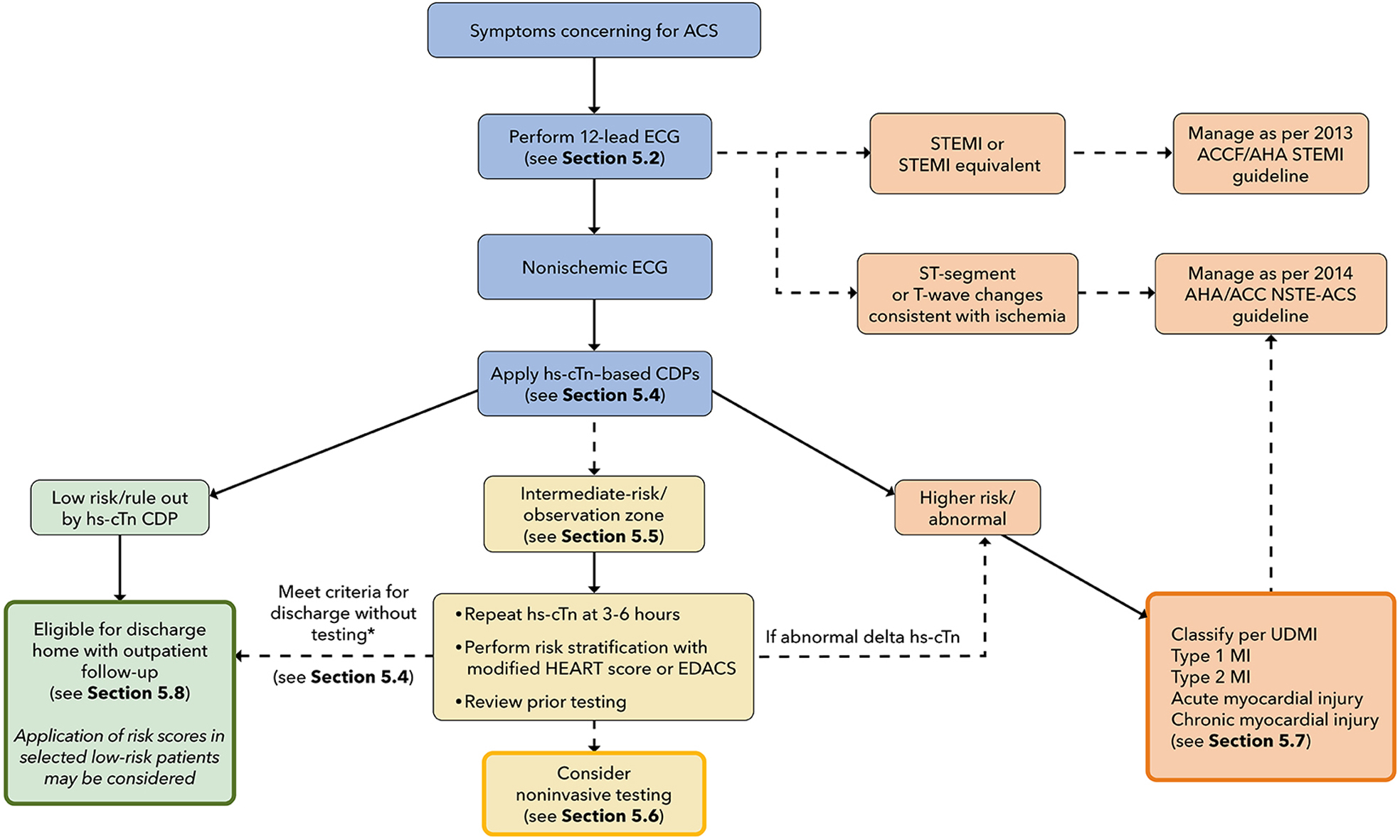
ACC 2022 chest pain evaluation pathway
Tap the figure to open it at full size

Initial Assessment

  • ECG within 10 minutes, with initial hs-cTn obtained promptly
  • Compare previous tracings when available
  • Persistent / recurrent symptoms or deterioration → repeat ECG
  • Evaluate ACS and other dangerous causes together; do not postpone the ECG while completing the rest of the chest-pain differential
  • Figure 1 — Pathway Summary: Kontos et al., ACC 2022, original Figure 1. Source
  • Figure note (*): Unchanged or minimally changing serial hs-cTn with reassuring context: normal coronary angiography within 2 years or stress testing within 1 year; symptoms inconsistent with ACS; chronic elevations matching prior values; or modified HEART ≤3 / EDACS <16

STEMI / Acute Coronary Occlusion Pattern

Ischemic ECG / High-Risk Clinical Findings

  • Ischemic ECG without STEMI: New / dynamic ischemic ST depression, T-wave inversion, transient ST elevation, or a Wellens pattern
  • Clinical instability: Shock, significant arrhythmia, acute heart failure, or refractory ischemic symptoms
  • High-risk evaluation → cardiology + monitored care + serial ECGs / hs-cTn
  • Do not use an accelerated discharge pathway
    • An initially normal troponin does not cancel an ischemic ECG
    • Serial troponins still help establish the diagnosis; they are not a prerequisite for recognizing high risk
  • Ongoing refractory ischemia / hemodynamic or electrical instability → urgent invasive assessment
  • References: ACC 2022 · ACC/AHA 2025.

Concerning / Equivocal ECG

  • Cardiologist review + serial ECGs ± urgent echo
  • Clarify potentially ischemic changes rather than assigning them to the nonischemic branch
  • Reference: ACC 2022.

Nonischemic ECG + Clinically Stable

  • Normal ECG / nonspecific ST-T changes; no convincing acute ischemic pattern
  • Eligible for the accelerated hs-cTn pathway when no clinical finding independently requires admission
  • Nonischemic ECG ≠ ACS excluded
  • Reference: ACC 2022.

hs-cTn Clinical Decision Pathway

Apply The Institutional Pathway

  • High-sensitivity cardiac troponin preferred
  • Use your institution’s assay, sampling times, cutoffs, and delta criteria
  • Common approaches include 0/1-hour, 0/2-hour, and selected single-draw rule-out
  • Interpret the initial value and change over time alongside the ECG, symptom timing, and clinical presentation
  • The pathway determines the next action: Rule-out / observation / further evaluation
    • These are management groups—not final diagnoses
  • Reference: ACC/AHA 2025.
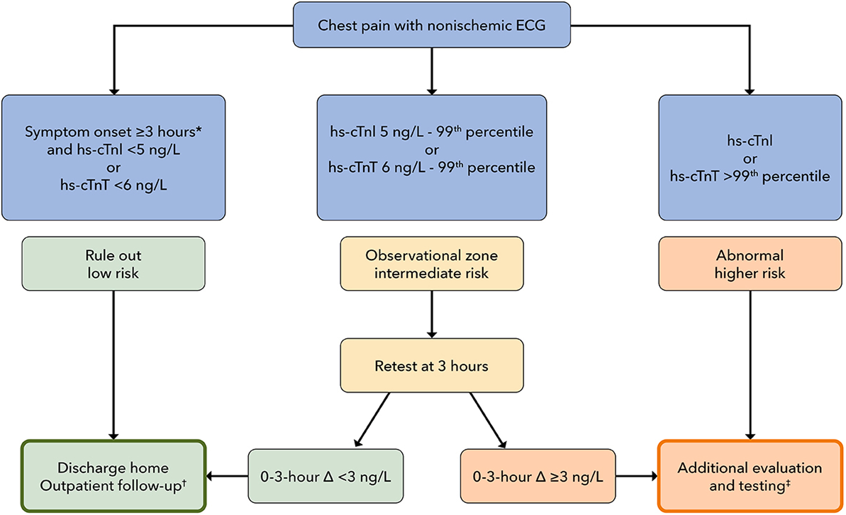
ACC 2022 High-STEACS early rule-out pathway
Tap the figure to open it at full size
  • Figure 2 — High-STEACS Early Rule-Out CDP: ACC 2022, original Figure 4. This is a specific pathway; use locally validated assays and thresholds. Source
  • Figure note (*): Trials accepted onset ≥2 hours before presentation; ACC recommends ≥3 hours between symptom onset and the first sample for immediate rule-out
  • Figure note (†): Arrange outpatient follow-up and selective testing; see source Sections 5.6 and 5.8
  • Figure note (‡): Additional evaluation may include observation / admission and anatomical or functional testing. Classify injury and treat acute MI appropriately; stable chronic injury may permit outpatient care. See Sections 5.6–5.8. The figure uses the 2022 MI terminology; the updated classification is described below

Selected Single-Draw Rule-Out

  • Nonischemic ECG + reassuring clinical assessment
  • Symptoms began ≥3 hours before the sample
  • hs-cTn below the institution’s validated very-low rule-out threshold
  • Not simply “one normal troponin”
    • Early / uncertain symptom onset or concerning findings require further assessment
  • Reference: ACC 2022.

Rule-Out / Low Risk

  • Nonischemic ECG + validated hs-cTn rule-out + low-risk clinical assessment
  • Discharge with follow-up when no other admission indication exists
  • No routine urgent stress test / coronary CTA / inpatient cardiac testing
  • HEART / EDACS: Not automatically required after a validated hs-cTn rule-out; use when incorporated into the institutional pathway or residual concern remains
  • Persistent concern for acute ischemia or another dangerous diagnosis requires further evaluation
  • References: AHA/ACC 2021 · ACC 2022.

Intermediate Risk / Observation

  • Modified HEART (HEAR; ACC 2022): History, ECG, age, and risk factors; 0–8 points. This version excludes troponin and must be paired with the appropriate troponin pathway
  • EDACS: Use the score within a validated clinical decision pathway; a score <16 alone is insufficient for discharge
  • Not ruled out; not clearly assigned to the abnormal / high-risk group
  • Repeat hs-cTn at 3–6 hours
  • Reassess symptoms / ECG; compare prior troponin values and review known CAD / previous cardiac testing
  • HEART / EDACS: May help refine disposition
  • Further testing: Selective coronary CTA / stress testing based on the remaining concern, known CAD, and prior results
  • Disposition: Selected patients may leave after reassuring reassessment with rapid follow-up; others need further observation or admission
  • Intermediate risk ≠ unstable angina: The evaluation is still incomplete
  • Reference: ACC 2022.

Abnormal / High Risk

  • Substantially elevated hs-cTn / significant rise or fall / other high-risk findings
  • Further evaluation; usually admission
  • Determine whether the abnormality represents MI or another cause of myocardial injury
  • An elevated troponin is not an automatic NSTEMI diagnosis or an instruction to give every patient the same ACS treatment
  • Reference: ACC 2022.

Diagnostic Interpretation

STEMI

  • Acute MI presenting with diagnostic ST-segment elevation
  • Immediate reperfusion pathway; do not wait for troponin
  • Acute coronary occlusion / STEMI-equivalent patterns: May require the same urgent pathway without meeting classic STEMI criteria
  • References: ACC/AHA 2025 · ACC 2022.

NSTEMI

  • Acute myocardial injury + evidence that the injury is due to myocardial ischemia, without a STEMI presentation
  • Acute injury: Troponin rise / fall with at least one value above the sex-specific 99th percentile
  • Ischemic evidence: Clinical presentation, ECG changes, and coronary / cardiac imaging when needed
  • An ischemic ECG is not required: NSTEMI can emerge from the initially nonischemic-ECG pathway
  • Reference: ACC/AHA 2025.
MI Mechanism
  • Primary: Acute coronary pathology
  • Secondary: Another acute illness causing myocardial oxygen supply–demand imbalance
  • Procedure-related: A cardiac-procedure complication
  • Classify the mechanism after establishing MI using the appropriate clinical and objective criteria
  • Troponin elevation during another illness alone does not establish secondary MI
  • Reference: Universal Definition 2026.

Unstable Angina

  • Convincing acute myocardial ischemia without acute troponin-defined myocardial injury
  • Serial testing helps establish the absence of acute injury
  • One negative troponin does not establish unstable angina
  • Negative serial troponins do not automatically establish it either: There must still be convincing evidence of acute ischemia
  • Reference: ACC/AHA 2025.

Myocardial Injury Without Acute MI

  • Acute injury: Troponin rise / fall with at least one value above the sex-specific 99th percentile, without sufficient evidence of infarction
  • Chronic injury suspected: Persistently elevated hs-cTn without a significant acute change; review prior values, clinical stability, and the underlying cause before confirming
  • Identify the cause rather than labeling every elevation NSTEMI
  • Reference: Universal Definition 2026.

No ACS Established

  • Consider another cardiac / noncardiac cause
  • MI rule-out does not exclude every cause of ischemic chest pain
  • A safe low-risk disposition does not require proving one particular benign diagnosis
  • Persistent concerning symptoms / findings → continue evaluation rather than forcing a reassuring label
  • Reference: AHA/ACC 2021.

In The Pit

  • Possible ACS → ECG first
    • STEMI / acute coronary occlusion pattern → immediate reperfusion
    • Ischemic ECG / high-risk clinical findings → cardiology + monitored care + serial hs-cTn
    • Concerning / equivocal ECG → cardiologist review + serial ECGs ± urgent echo
    • Nonischemic ECG + clinically stable → institutional hs-cTn pathway
  • hs-cTn pathway → next step
    • Rule-out / low risk → discharge when the complete assessment supports it
    • Intermediate → repeat hs-cTn + reassess ± HEART / EDACS ± coronary CTA / stress testing
    • Abnormal / high risk → further evaluation / usually admission; classify myocardial injury
  • ECG + serial hs-cTn + clinical findings → diagnostic interpretation
    • Acute myocardial injury + ischemia → MI: STEMI / NSTEMI; classify the mechanism
    • Convincing acute ischemia without acute myocardial injury → unstable angina
    • Troponin elevation without acute MI → acute / chronic myocardial injury
    • No ACS established → evaluate other causes / determine disposition
    • Negative troponins alone → neither an unstable-angina diagnosis nor automatic discharge

04Other Causes Of Chest Pain

  • Emergent causes addressed / ACS risk assessed → evaluate other cardiac and noncardiac causes. A low-risk ACS assessment does not exclude another dangerous diagnosis.

Stable Angina

  • Consider: A reproducible exertional pattern without a new or worsening acute pattern
  • Acute change matters: New rest pain, increasing frequency / severity, or a concerning ECG / troponin pattern returns the patient to ACS evaluation
  • Disposition: Arrange appropriate follow-up and selective further evaluation based on known CAD, symptoms, and prior testing AHA/ACC 2021

Differential

  • GI / esophageal: GERD, esophagitis / esophageal spasm, gastritis / PUD, biliary disease, pancreatitis → see Abdominal Pain — Upper.
  • Chest wall / musculoskeletal: Costochondritis, muscle strain, rib injury. Reproducible tenderness supports a chest-wall source but does not independently exclude ACS.
  • Pulmonary / pleural: Pneumonia, bronchitis, pleurisy.
  • Skin / neurologic: Herpes zoster / cervical radiculopathy.
  • Anxiety / panic / hyperventilation: Assess in context—not simply because initial testing is negative.

Before Discharge

  • Reassess symptoms, vital signs, and examination. Persistent concerning findings → further evaluation; otherwise provide symptom-directed care, follow-up, and return precautions.
Reference

Must-Read References

View Full References

Additional Cited Guidance

Chest Pain / Pulmonary Causes

Aortic Dissection

Pericarditis / Myocarditis

Esophageal Perforation

Clinical Decision Tools

Privacy & analytics choices