The Algorithm...
- Sudden weakness, speech / visual change, or severe new gait / coordination difficulty → assess for acute stroke. Stabilization, imaging, and reperfusion assessment proceed together.
01 Initial Evaluation
- Activate the stroke team / telestroke; establish IV access and monitoring.
- ABCs + Glucose: Support airway / ventilation; correct hypoglycemia, hypoxemia, and hypotension.
- Establish last known well, symptom discovery, baseline function, and anticoagulant / last dose. See Initial Evaluation.
02 Stroke Syndrome / Mimic?
- Define the deficit: language, vision, gaze, face, strength, sensation, coordination, and neglect.
- NIHSS + What Can The Patient No Longer Do? Low NIHSS does not mean nondisabling. Correct plausible reversible causes without exhaustive mimic testing.
- Resolved symptoms still require ischemia assessment; a negative MRI does not independently settle TIA versus stroke. See Clinical Assessment.
03 Urgent Imaging
- Noncontrast CT → Hemorrhage? If present, follow the Intracranial Hemorrhage pathway.
- CTA Head / Neck → Occlusion Or Dissection? Obtain promptly when indicated.
- Normal CT does not exclude ischemic stroke. Use MRI / perfusion for selected diagnostic or extended-window questions without delaying indicated early IVT. See Imaging.
04 Reperfusion Candidate?
- Assess IV Thrombolysis (IVT) And Endovascular Thrombectomy (EVT) Together—Not One After The Other.
- Disabling Ischemic Deficit: Standard IVT window ≤4.5 hours; complete the safety / BP assessment.
- Occlusion Suitable For Thrombectomy: Immediate specialist assessment / transfer; selected patients qualify through 24 hours. Do not wait for IVT response.
- Wake-Up / Late Presentation: Imaging-based selection may permit treatment. Truly nondisabling deficits require continued evaluation and an appropriate antiplatelet pathway. See Reperfusion.
05 Other Immediate Care
- BP: Use the target for IVT / thrombectomy / neither; correct hypotension.
- Antiplatelets: After hemorrhage exclusion and the reperfusion decision; selected minor noncardioembolic stroke / high-risk TIA → short-course dual antiplatelet therapy (DAPT).
- After IVT: Generally withhold antithrombotics for 24 hours and obtain follow-up imaging before starting.
- Swallowing: Nothing by mouth until swallowing safety is assessed; treat significant glucose abnormalities and fever. See Immediate Care.
06 Reassess / Disposition
- Repeat the deficit, consciousness, BP, glucose, and breathing. Deterioration → immediate reassessment / imaging.
- Discharge: Selected TIA after appropriate initial assessment, prevention, and a secured rapid follow-up pathway.
- Monitored Admission: Stroke, high-risk TIA, or required post-reperfusion monitoring.
- ICU Admission: Airway / hemodynamic support, major treatment complications, or neurologic deterioration. See Disposition.
Initial Evaluation
ABCs / Glucose / Monitoring
- Stroke Activation: Involve the stroke team / telestroke service; obtain IV access, cardiac monitoring, bedside glucose, and rapid imaging.
- Airway: Assess consciousness, bulbar function, ventilation, and airway protection. Intubate when these fail; GCS alone is not the decision.
- Oxygenation: Treat hypoxemia to maintain SpO₂ >94%. Routine oxygen for every normoxic patient is unnecessary; selected thrombectomy protocols are separate. AHA/ASA 2026 — Airway / Oxygenation.
- Glucose / Perfusion: Correct hypoglycemia, hypotension, and clinically important volume depletion; repeat the examination after correction. AHA/ASA 2026 — Supportive Care.
Last Known Well / Baseline
- Record last known well, symptom discovery, and witnessed onset separately. Obtain the witness's account and contact information.
- Wake-up symptoms: Awakening is not last known well. Document fluctuations and any convincing interval of complete recovery; partial improvement does not reset the clock.
- Review baseline independence, mobility, cognition, communication, and existing deficits.
- Review anticoagulants / last doses, antiplatelets, recent bleeding, surgery, trauma, and intracranial disease. Baseline disability requires individualized assessment rather than automatic exclusion. AHA/ASA 2026 — Eligibility.
Stroke Syndrome / Mimic?
History / Examination
- Hemispheric: Unilateral weakness / numbness, aphasia, neglect, gaze preference, or visual-field loss.
- Deep / Lacunar: Pure motor, pure sensory, or sensorimotor deficits may arise from subcortical injury. Combined motor and sensory loss does not establish a cortical lesion.
- Posterior Circulation: Diplopia, dysarthria, dysphagia, abnormal eye movements, crossed findings, severe gait / truncal ataxia, or reduced consciousness. Vertigo may dominate.
- Dissection: New headache / neck pain, painful Horner syndrome, or relevant trauma. Chest / back pain with pulse or perfusion abnormalities raises concern for aortic disease.
- Endocarditis: Fever, bacteremia risk, embolic findings, or a concerning murmur. This changes reperfusion safety as well as the differential. AHA/ASA 2026 — Special Circumstances.
- Examine language, gaze, visual fields, face, limb strength / sensation, coordination, and neglect; assess gait when safe. Clinical localization directs imaging but does not replace it. AHA 2023 — Clinical Assessment.
NIHSS / What Can The Patient No Longer Do?
- Document NIHSS at baseline and after treatment or clinical change.
- Low NIHSS can still be disabling: Aphasia, visual loss, or inability to walk may prevent usual activities despite a low total score.
- Assess communication, ambulation, self-care, and usual work. Improvement that leaves a disabling deficit is not complete recovery. AHA/ASA 2026 — Disability.
TIA Versus Ischemic Stroke
- TIA: Transient focal ischemic dysfunction with complete recovery and no demonstrated acute infarction.
- Ischemic Stroke: Neurologic dysfunction caused by focal infarction; symptoms may persist or resolve. A relevant acute infarct establishes stroke even after full recovery. AHA/ASA — Definitions.
- Apply the following distinctions when the clinical event is most consistent with ischemia—not solely because focal symptoms occurred.
| Clinical Course | MRI Finding | Interpretation |
|---|---|---|
| Focal symptoms resolve completely after a few hours | No acute infarct demonstrated | Clinical TIA, when ischemia is the best explanation |
| Focal symptoms resolve completely | Relevant acute infarct | Ischemic stroke, despite recovery |
| Focal deficit persists | Relevant acute infarct | Ischemic stroke |
| Focal deficit persists | Initial MRI negative | Suspected ischemic stroke, including DWI-negative stroke; continue evaluation |
- Bedside Example: Three hours of hemiparesis, full recovery, and negative DWI generally fits TIA when the history supports ischemia. The same episode with a corresponding acute infarct is stroke. AHA 2023 — TIA Definition.
- Negative MRI is not proof that no infarction occurred. Small, early, and posterior-circulation infarcts may be missed; symptoms and trajectory still matter. See MRI Limitations. Edlow 2017.
- Persistent focal ischemic deficits ≥24 hours, with other causes excluded, can establish clinical evidence of infarction despite negative imaging. Do not wait 24 hours to diagnose suspected stroke or assess reperfusion; this is a classification criterion, not a treatment delay. AHA/ASA — Clinical Definition.
Important Mimics
- Hypoglycemia: Correct and reassess; persistent focal deficits still require stroke evaluation.
- Seizure / Postictal Weakness: Assess the witnessed sequence and recovery. Seizure at onset does not prove that the remaining deficit is postictal.
- Migraine Aura: Prior identical episodes and spreading symptoms support the diagnosis; a first or atypical event requires further assessment.
- Other Causes: Toxic / metabolic disturbance, hypertensive encephalopathy, infection, or mass lesion when supported by the presentation.
- Functional Symptoms: Require positive clinical findings—not a psychiatric history or normal CT alone.
- Do not delay eligible treatment for exhaustive mimic testing; resolve material uncertainty with the stroke team. AHA/ASA 2026 — Diagnostic Uncertainty.
Urgent Imaging
Noncontrast Head CT
- Exclude hemorrhage, assess established ischemic injury, and identify major alternatives.
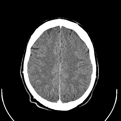
- Early ischemic findings include loss of gray–white differentiation, sulcal effacement, or a hyperdense artery. A normal CT does not exclude acute ischemic stroke.
- ASPECTS describes early anterior-circulation injury; interpret it within the reperfusion pathway rather than as a universal exclusion. AHA/ASA 2026 — Imaging.
CTA Head / Neck
- Identify large-vessel occlusion, relevant stenosis, or dissection. Contact the thrombectomy team promptly to determine whether the occlusion is suitable for treatment.
- Suspected Large-Vessel Occlusion (LVO): Do not delay urgent CTA / CT perfusion for a creatinine result. Address a known severe contrast reaction through the emergency imaging pathway.
- Coordinate imaging with IV thrombolysis preparation; additional studies should not delay otherwise indicated early treatment. AHA/ASA 2026 — Vascular Imaging.
MRI / Perfusion Imaging
- MRI-DWI: Infarct detection when CT is unrevealing or the diagnosis remains uncertain.
- DWI–FLAIR / Perfusion Mismatch: Selected unknown-onset or extended-window eligibility. Follow the specific imaging criteria. See Extended Windows.
- Persistent Deficit Despite Negative DWI: Continue stroke assessment, review the vascular imaging, and discuss repeat MRI when appropriate. Do not relabel an ongoing deficit as TIA merely because MRI is negative. Edlow 2017.
Evidence — MRI-Negative Stroke: A meta-analysis found negative DWI in approximately 6.8% of clinically diagnosed acute ischemic strokes, with higher odds in posterior-circulation disease. The proportion varies with the population and imaging timing; it is not a universal MRI miss rate. Edlow 2017.
Laboratory / Cardiac Testing
- Confirm glucose before IV thrombolysis. Obtain CBC / platelets, electrolytes / renal function, and relevant coagulation studies.
- Routine platelet / coagulation results need not delay IVT when no abnormality is suspected; anticoagulant exposure or bleeding history changes the assessment. AHA/ASA 2026 — Laboratory Timing.
- Obtain ECG and troponin without delaying reperfusion; additional testing follows the suspected cause. AHA/ASA 2026 — Cardiac Testing.
Reperfusion Candidate?
- Assess IV thrombolysis (IVT) and endovascular thrombectomy (EVT) in parallel. Eligible patients may need both; do not wait for thrombolytic response before thrombectomy / transfer. AHA/ASA 2026 — Combined Treatment · ACEP 2024.
IV Thrombolysis
- Within 4.5 Hours: Treat eligible disabling ischemic stroke promptly after hemorrhage exclusion and safety assessment.
- Explain potential functional benefit and serious bleeding risk to the patient / surrogate when feasible; document the decision without avoidable delay.
- Age over 80, a low NIHSS, or partial improvement does not independently exclude treatment. A truly nondisabling presentation follows a different pathway. AHA/ASA 2026 — IVT Decisions.
Evidence — Alteplase: Outcomes with no significant disability at three to six months occurred in 32.9% versus 23.1% with alteplase versus control within three hours, and 35.3% versus 30.1% at three to 4.5 hours. Early fatal intracranial hemorrhage increased; a 90-day mortality benefit was not established. These are trial averages, not individual predictions. Emberson 2014.
Agent / Dose
| Agent | Adult Ischemic-Stroke Dose | Administration |
|---|---|---|
| Tenecteplase | 0.25 mg/kg IV; maximum 25 mg | Single bolus using the stroke-specific preparation / weight-based protocol |
| Alteplase | 0.9 mg/kg IV; maximum 90 mg | 10% over one minute; remaining 90% over 60 minutes |
- Confirm weight, concentration, and total dose. Do not substitute STEMI tenecteplase dosing. AHA/ASA 2026 — Administration.
Evidence — AcT: Tenecteplase was noninferior to alteplase in eligible disabling stroke. Single-bolus administration is practical; the trial did not establish universal superiority. Menon 2022.
Bleeding Risk / Eligibility
- Complete the current stroke-team checklist; the following are major safety checks, not an exhaustive contraindication table.
- Hemorrhage / Major Bleeding: Exclude intracranial hemorrhage, suspected SAH, active major bleeding, aortic dissection, and infective endocarditis before IVT.
- Imaging / Recent Injury: Assess extensive established hypodensity and review intracranial / spinal surgery, significant head trauma, recent stroke, and intracranial lesions using the specific eligibility criteria. AHA/ASA 2026 — Eligibility.
- Coagulation: Platelets <100,000/µL, INR >1.7, or clinically important coagulation abnormalities preclude standard IVT. Interpret tests according to the drug exposure. AHA/ASA 2026 — Coagulopathy.
- BP: Must remain below the pretreatment threshold. See Blood Pressure.
- Pregnancy, prior ICH, baseline disability, and other complex circumstances require individualized assessment. An IVT exclusion does not automatically exclude thrombectomy. AHA/ASA 2026 — Individualized Decisions.
Anticoagulant Exposure
- Identify the agent, last dose, and renal function: warfarin, heparin, LMWH, and direct oral anticoagulants (DOACs) require different assessment.
- A normal INR does not exclude clinically important DOAC activity. Use drug-appropriate testing when available.
- Recent DOAC exposure, particularly within 48 hours, requires specialist risk–benefit assessment; IVT safety remains uncertain. Reversal solely to permit IVT is not an automatic step. AHA/ASA 2026 — DOACs.
Unknown Onset / Extended Windows
- Unknown Onset: Selected DWI–FLAIR mismatch may support IVT within 4.5 hours of symptom recognition.
- 4.5–9 Hours / Wake-Up Stroke: In patients not eligible for EVT, automated perfusion selection may support IVT at 4.5–9 hours from last known well, or within nine hours of the midpoint of sleep for qualifying wake-up presentations.
- 4.5–24 Hours With LVO: Selected salvageable-tissue cases in which thrombectomy cannot be performed may qualify for specialist-directed IVT. This is not a universal 24-hour thrombolysis window. AHA/ASA 2026 — Extended Windows · July 2026 Correction.
Evidence — WAKE-UP: MRI-selected alteplase improved functional outcomes in unknown-onset stroke; planned thrombectomy patients were excluded. Thomalla 2018.
Endovascular Thrombectomy
- Proximal Anterior Occlusion: Intracranial ICA / proximal MCA → immediate assessment; selected patients qualify through 24 hours.
- Large Core: Substantial established injury is not an automatic exclusion. Apply current imaging, timing, and baseline-function criteria.
- Basilar Occlusion: Urgent assessment within 24 hours; evidence is strongest in selected patients with NIHSS ≥10 and suitable imaging / baseline function. Lower scores require individualized assessment.
- Medium / Distal Occlusion: Do not extend proximal-vessel benefit to every clot. Selected dominant M2 disease differs from routine nondominant / distal occlusion treatment. AHA/ASA 2026 — EVT Selection.
- Transfer: Send imaging, timeline, examination, anticoagulant history, treatment doses / times, and BP trend. Do not wait for the alteplase infusion to finish.
Evidence — HERMES: NNT 2.6 for improvement by at least one modified Rankin Scale category—not one death prevented or one additional independent survivor. Goyal 2016.
Other Immediate Care
Blood Pressure — IVT, Thrombectomy, Or Neither?
| Clinical Situation | BP Instruction — mm Hg |
|---|---|
| Before IVT | Lower to SBP <185 and DBP <110; maintain control before treatment |
| After IVT | Maintain <180/105 for at least 24 hours |
| Before EVT Without IVT | ≤185/110 is reasonable |
| During / For 24 Hours After EVT | Guideline ceiling ≤180/105; obtain the treating team's specific target |
| No IVT / EVT; BP <220/120 | Usually leave BP untreated acutely unless another emergency requires reduction |
| No IVT / EVT; SBP ≥220 Or DBP ≥120 | Individualized, cautious reduction; avoid abrupt drops. Outcome benefit remains uncertain |
- 220/120 is a threshold for considering intervention—not a treatment target or required minimum. Correct hypotension and assess new deficits during BP reduction.
- ACS, acute heart failure, aortic dissection, or another hypertensive emergency changes the plan. Do not automatically restart antihypertensives at 24 hours regardless of the clinical course. AHA/ASA 2026 — BP.
- After Successful EVT: Do not routinely force SBP below 140. Avoid hypotension and excessive variability; follow the specialist's reperfusion-specific instructions. AHA/ASA 2026 — Post-EVT BP.
Evidence — Post-EVT BP: OPTIMAL-BP found worse functional outcomes with a uniform SBP target below 140. HOPE found better recovery with reperfusion-guided targets in selected patients. Different populations, exclusions, and protocols do not establish one universally appropriate lower target. OPTIMAL-BP 2023 · HOPE 2026.
BP Medications
- Labetalol: 10–20 mg IV over one to two minutes; may repeat once when appropriate for heart rate / cardiopulmonary status.
- Nicardipine: 5 mg/hour IV, increasing by 2.5 mg/hour every 5–15 minutes, maximum 15 mg/hour. Titrate to the applicable target and repeat the neurologic examination. Stroke Nebraska — BP Regimens.
Swallowing / Glucose / Temperature
- Swallowing: Nothing by mouth, including oral medication, until swallowing safety is assessed; use an appropriate nonoral route when needed. AHA/ASA 2026 — Dysphagia.
- Glucose: Correct hypoglycemia; for persistent hyperglycemia, 140–180 mg/dL is reasonable with close monitoring.
- Fever: Treat and assess for infection or another cause. AHA/ASA 2026 — Glucose / Temperature.
Evidence — SHINE: Intensive glucose control did not improve function and increased severe hypoglycemia. Johnston 2019.
Antiplatelets / Anticoagulation
- Aspirin: After hemorrhage exclusion and the reperfusion decision, start within 24–48 hours when appropriate; initial oral dosing commonly 160–325 mg, with a nonoral route when swallowing is unsafe. Aspirin does not replace indicated reperfusion. AHA/ASA 2026 — Antiplatelets.
- Dual Antiplatelet Therapy (DAPT): Selected minor noncardioembolic stroke (NIHSS ≤3) or high-risk TIA (ABCD2 ≥4), without IVT and with acceptable bleeding risk → aspirin + clopidogrel early, preferably within 24 hours, for 21 days, then single-agent therapy.
- Use the protocol-specified loading regimen; usual maintenance is aspirin 81 mg daily + clopidogrel 75 mg daily. Record the transition date. Expanded eligibility in selected atherosclerotic disease requires its specific criteria. AHA/ASA 2026 — DAPT.
- After IVT: Generally withhold antithrombotics until 24 hours after treatment, with follow-up CT / MRI before initiation; exceptional indications require specialist direction. AHA/ASA 2026 — Post-IVT Care.
- Anticoagulation: Therapeutic heparin is not routine acute arterial-stroke treatment. AF-related secondary prevention requires a separate timing decision based on infarct burden, hemorrhage risk, and treatment; DAPT is not an equivalent substitute. AHA/ASA 2026.
- ABCD2 is used here to describe a studied treatment population—not to decide discharge on its own.
Evidence — DAPT: CHANCE/POINT found most benefit within 21 days. ARAMIS supported DAPT in selected minor, nondisabling stroke; it does not support withholding IVT from every low-NIHSS patient. Pan 2019 · ARAMIS 2023.
Determine The Cause
- Cardioembolic: ECG / telemetry; selected echo for structural disease, thrombus, or endocarditis.
- Large-Artery: Review head / neck imaging for relevant stenosis or dissection; symptomatic carotid disease needs urgent specialist assessment.
- Small-Vessel: Match the syndrome to imaging; avoid assuming the mechanism from symptoms alone.
- Connect the cause to antithrombotic choice, lipid therapy, risk-factor treatment, and follow-up. Broad etiologic testing must not delay reperfusion. AHA 2023 — Mechanism / Prevention.
TIA — Urgent Evaluation / Prevention
- Obtain brain / vascular assessment and ECG; MRI-DWI ideally within 24 hours. When MRI is unavailable, arrange timely imaging through observation, admission, transfer, or an established outpatient pathway.
- Assess relevant stenosis, dissection, AF, and other supported mechanisms. Begin appropriate prevention once hemorrhage and treatment conflicts are excluded.
- Low ABCD2 or negative DWI does not establish safe discharge. Recurrent events, abnormal vascular imaging, and follow-up access change the plan.
- A relevant acute infarct despite symptom resolution is ischemic stroke, not “TIA with infarction.” Recurrent active deficits re-enter the stroke pathway. See TIA Versus Stroke and Disposition. AHA 2023 — TIA Assessment.
Reassess / Disposition
Response / Complications
- Repeat the deficit, consciousness, BP, glucose, and breathing. Confirm outstanding reperfusion decisions and support needs.
- After IVT: Neurologic / BP checks every 15 minutes for two hours, every 30 minutes for six hours, then hourly through 24 hours. Defer unnecessary invasive procedures when safe. AHA/ASA 2026 — Monitoring.
- New Headache / Vomiting / Neurologic Worsening: Emergency CT and stroke-team reassessment; stop ongoing alteplase. Tenecteplase has already been administered as a bolus.
- Suspected Thrombolytic Bleeding: CBC, coagulation studies, fibrinogen, type / cross, and immediate reversal-protocol / specialist activation. AHA/ASA 2026 — Bleeding.
- Tongue / Oropharyngeal Swelling: Assess progression, summon airway expertise, and stop ongoing alteplase. Follow the thrombolytic-angioedema pathway. AHA/ASA 2026 — Angioedema.
- Declining Consciousness / Large Infarct: Assess edema, mass effect, or hydrocephalus; urgent neurocritical / neurosurgical evaluation. See the separate Intracranial Hemorrhage and Airway pathways.
Discharge
- Selected TIA presentations with sustained resolution, reassuring completed initial assessment, prevention started, and a secured rapid pathway.
- Arrange indicated testing and a definite TIA / neurology appointment, preferably within 48 hours. Confirm medication access, transport, and ability to return; recurrent focal symptoms require immediate EMS activation. AHA 2023 — Disposition.
Monitored Admission
- Ischemic stroke requiring stroke-unit assessment, telemetry, neurologic surveillance, swallowing / rehabilitation assessment, or urgent investigation.
- Acute infarction despite resolved symptoms: Ischemic stroke requiring monitored stroke assessment—not a TIA discharge branch.
- TIA: Recurrent symptoms, relevant stenosis, concerning cardiac findings, or an unsafe outpatient plan. Structured observation may complete evaluation in selected patients.
- Post-IVT care requires a stroke unit or ICU capable of the mandated monitoring. Transfer immediately when necessary reperfusion / stroke care is unavailable. AHA/ASA 2026 — Stroke Care.
ICU Admission
- Airway / ventilatory failure, hemodynamic instability, or intensive titrated support.
- Symptomatic intracranial hemorrhage, major treatment-related bleeding, or airway-threatening angioedema.
- Deteriorating neurologic status, substantial edema / mass effect, hydrocephalus, or need for neurocritical / neurosurgical access. Level of care follows support and monitoring needs—not the treatment label alone. AHA/ASA 2026.
In The Pit
- Unstable / Deteriorating: Support airway and perfusion; check glucose. New worsening after IVT → emergency CT + stroke team; stop ongoing alteplase. See Complications.
- Sudden Focal Deficit: Stroke activation + last known well + NIHSS / disability + urgent CT ± CTA. A normal CT does not clear the patient.
- Disabling Deficit: Assess IVT immediately; aphasia, visual loss, or inability to walk may be disabling despite low NIHSS. Partial improvement is not full recovery.
- Large-Vessel Occlusion: Thrombectomy assessment / transfer in parallel. Do not wait to see whether IVT works.
- Persistent But Nondisabling Deficit: Continue stroke evaluation and prevention; selected noncardioembolic minor stroke → DAPT, not automatic IVT. Negative MRI alone does not convert an ongoing deficit into TIA.
- Symptoms Resolved: Urgent TIA evaluation; relevant acute infarct = stroke. Negative MRI with complete recovery can fit TIA; discharge still requires appropriate assessment and a secured rapid follow-up plan. See Disposition.
Related Algorithms
Pop Quiz Hotshot
 Pop Quiz HotshotAcute Stroke10 QuestionsStart Quiz
Must-Read References
View Full References
- Emberson Et Al., 2014 — Alteplase Individual-Patient Meta-Analysis. Effect of Treatment Delay, Age, and Stroke Severity on the Effects of Intravenous Thrombolysis With Alteplase for Acute Ischaemic Stroke. Lancet. Time-dependent functional benefit and hemorrhage risk.
- Goyal Et Al., 2016 — HERMES. Endovascular Thrombectomy After Large-Vessel Ischaemic Stroke: A Meta-Analysis of Individual Patient Data From Five Randomised Trials. Lancet. Foundational disability benefit; interpret the NNT by its actual endpoint.
- Menon Et Al., 2022 — AcT. Intravenous Tenecteplase Compared With Alteplase for Acute Ischaemic Stroke in Canada. Lancet. Randomized noninferiority evidence in eligible disabling stroke.
- Thomalla Et Al., 2018 — WAKE-UP. MRI-Guided Thrombolysis for Stroke With Unknown Time of Onset. New England Journal of Medicine. DWI–FLAIR selection; planned thrombectomy was excluded.
- AHA/ASA, 2026 — Acute Ischemic Stroke Guideline. 2026 Guideline for the Early Management of Patients With Acute Ischemic Stroke. Stroke. Main U.S. framework, with the July 2026 Correction · EVT Selection Algorithm.
- ACEP, 2024 — Thrombolysis Clinical Policy. Clinical Policy: Use of Thrombolytics for the Management of Acute Ischemic Stroke in the Emergency Department. Annals of Emergency Medicine. IVT before thrombectomy and shared decision-making.
- Amin Et Al., 2023 — AHA TIA Statement. Diagnosis, Workup, Risk Reduction of Transient Ischemic Attack in the Emergency Department Setting. Stroke. Imaging, prevention, and resource-dependent disposition.
- Sacco Et Al., 2013 — AHA/ASA Stroke Definitions. An Updated Definition of Stroke for the 21st Century. Stroke. Tissue-based classification and clinical evidence of infarction when imaging is negative.
Additional Cited Guidance
- Edlow Et Al., 2017. Diagnosis of DWI-Negative Acute Ischemic Stroke: A Meta-Analysis. Neurology. Imaging limits, particularly in posterior-circulation disease.
- Chen Et Al., 2023 — ARAMIS. Dual Antiplatelet Therapy vs Alteplase for Patients With Minor Nondisabling Acute Ischemic Stroke. JAMA. Selected nondisabling population—not all low-NIHSS strokes.
- Pan Et Al., 2019 — CHANCE/POINT Pooled Analysis. Outcomes Associated With Clopidogrel-Aspirin Use in Minor Stroke or Transient Ischemic Attack. JAMA Neurology. Most DAPT benefit occurred within 21 days.
- Nam Et Al., 2023 — OPTIMAL-BP. Intensive vs Conventional Blood Pressure Lowering After Endovascular Thrombectomy in Acute Ischemic Stroke. JAMA. Harm from uniform intensive post-EVT reduction.
- Camps-Renom Et Al., 2026 — HOPE. Personalized Blood Pressure Targeting After Endovascular Therapy for Acute Ischemic Stroke. JAMA Neurology. Reperfusion-guided targets in a selected population.
- Johnston Et Al., 2019 — SHINE. Intensive vs Standard Treatment of Hyperglycemia and Functional Outcome in Patients With Acute Ischemic Stroke. JAMA. No functional advantage and more severe hypoglycemia with intensive treatment.
- Stroke Nebraska, 2024 — Acute Stroke Treatment Protocol. Practical BP-drug regimens; eligibility follows the newer AHA/ASA guidance above.